• 文献检索
  • 文档翻译
  • 深度研究
  • 学术资讯
  • Suppr Zotero 插件Zotero 插件
  • 邀请有礼
  • 套餐&价格
  • 历史记录
应用&插件
Suppr Zotero 插件Zotero 插件浏览器插件Mac 客户端Windows 客户端微信小程序
定价
高级版会员购买积分包购买API积分包
服务
文献检索文档翻译深度研究API 文档MCP 服务
关于我们
关于 Suppr公司介绍联系我们用户协议隐私条款
关注我们

Suppr 超能文献

核心技术专利:CN118964589B侵权必究
粤ICP备2023148730 号-1Suppr @ 2026

文献检索

告别复杂PubMed语法,用中文像聊天一样搜索,搜遍4000万医学文献。AI智能推荐,让科研检索更轻松。

立即免费搜索

文件翻译

保留排版,准确专业,支持PDF/Word/PPT等文件格式,支持 12+语言互译。

免费翻译文档

深度研究

AI帮你快速写综述,25分钟生成高质量综述,智能提取关键信息,辅助科研写作。

立即免费体验

用于示踪剂动力学测量的动态心肌灌注体模模型的开发。

Development of a dynamic myocardial perfusion phantom model for tracer kinetic measurements.

作者信息

Kamphuis Marije E, Kuipers Henny, Verschoor Jacqueline, van Hespen Johannes C G, Greuter Marcel J W, Slart Riemer H J A, Slump Cornelis H

机构信息

Multi-Modality Medical Imaging (M3i) Group, Faculty of Science and Technology, Technical Medical Centre, University of Twente, P.O. Box 217, 7500 AE, Enschede, The Netherlands.

Robotics and Mechatronics (RaM) Group, Faculty of Electrical Engineering Mathematics and Computer Science, University of Twente, Enschede, The Netherlands.

出版信息

EJNMMI Phys. 2022 Apr 25;9(1):31. doi: 10.1186/s40658-022-00458-y.

DOI:10.1186/s40658-022-00458-y
PMID:35467161
原文链接:https://pmc.ncbi.nlm.nih.gov/articles/PMC9038974/
Abstract

BACKGROUND

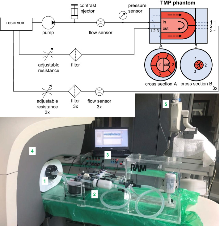
Absolute myocardial perfusion imaging (MPI) is beneficial in the diagnosis and prognosis of patients with suspected or known coronary artery disease. However, validation and standardization of perfusion estimates across centers is needed to ensure safe and adequate integration into the clinical workflow. Physical myocardial perfusion models can contribute to this clinical need as these can provide ground-truth validation of perfusion estimates in a simplified, though controlled setup. This work presents the design and realization of such a myocardial perfusion phantom and highlights initial performance testing of the overall phantom setup using dynamic single photon emission computed tomography.

RESULTS

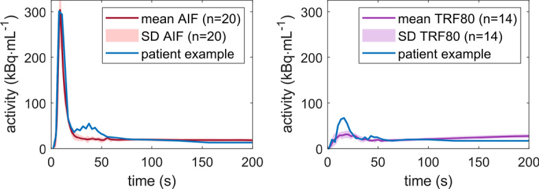
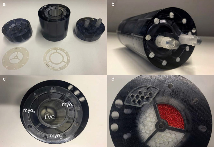
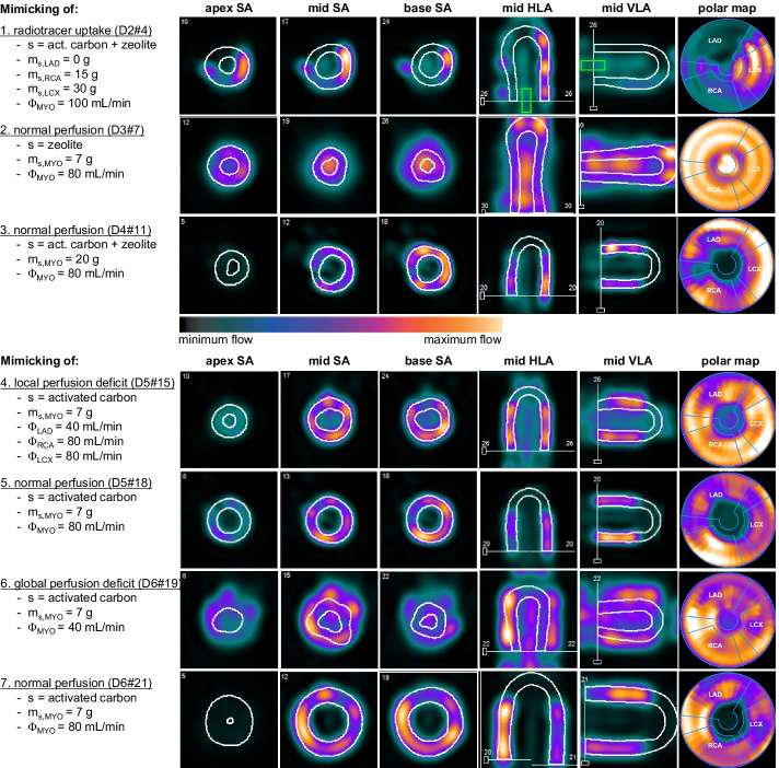
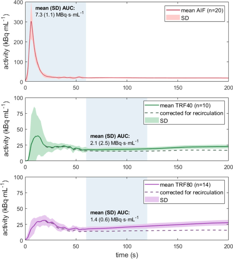
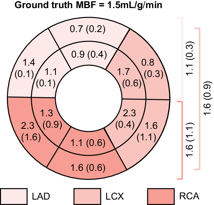
Due to anatomical and (patho-)physiological representation in the 3D printed myocardial perfusion phantom, we were able to acquire 22 dynamic MPI datasets in which Tc-labelled tracer kinetics was measured and analyzed using clinical MPI software. After phantom setup optimization, time activity curve analysis was executed for measurements with normal myocardial perfusion settings (1.5 mL/g/min) and with settings containing a regional or global perfusion deficit (0.8 mL/g/min). In these measurements, a specific amount of activated carbon was used to adsorb radiotracer in the simulated myocardial tissue. Such mimicking of myocardial tracer uptake and retention over time satisfactorily matched patient tracer kinetics. For normal perfusion levels, the absolute mean error between computed myocardial blood flow and ground-truth flow settings ranged between 0.1 and 0.4 mL/g/min.

CONCLUSION

The presented myocardial perfusion phantom is a first step toward ground-truth validation of multimodal, absolute MPI applications in the clinical setting. Its dedicated and 3D printed design enables tracer kinetic measurement, including time activity curve and potentially compartmental myocardial blood flow analysis.

摘要

背景

绝对心肌灌注成像(MPI)对疑似或已知冠心病患者的诊断和预后有益。然而,需要对各中心的灌注估计进行验证和标准化,以确保安全且充分地融入临床工作流程。物理心肌灌注模型可满足这一临床需求,因为它们能在简化但可控的设置中为灌注估计提供真实的验证。本文介绍了这种心肌灌注体模的设计与实现,并重点展示了使用动态单光子发射计算机断层扫描对整个体模设置进行的初始性能测试。

结果

由于3D打印的心肌灌注体模具有解剖学和(病理)生理学表现,我们能够获取22个动态MPI数据集,其中使用临床MPI软件测量和分析了锝标记示踪剂的动力学。在优化体模设置后,对正常心肌灌注设置(1.5 mL/g/min)以及包含局部或整体灌注不足的设置(0.8 mL/g/min)进行了时间-活度曲线分析。在这些测量中,使用特定量的活性炭在模拟心肌组织中吸附放射性示踪剂。这种对心肌示踪剂摄取和保留随时间变化的模拟与患者示踪剂动力学令人满意地匹配。对于正常灌注水平,计算得到的心肌血流量与真实血流设置之间的绝对平均误差在0.1至0.4 mL/g/min之间。

结论

所呈现的心肌灌注体模是在临床环境中对多模态绝对MPI应用进行真实验证的第一步。其专门的3D打印设计能够进行示踪剂动力学测量,包括时间-活度曲线以及潜在的房室心肌血流量分析。

https://cdn.ncbi.nlm.nih.gov/pmc/blobs/19c2/9038974/b6f019fb514e/40658_2022_458_Fig7_HTML.jpg
https://cdn.ncbi.nlm.nih.gov/pmc/blobs/19c2/9038974/74c235caa856/40658_2022_458_Fig1_HTML.jpg
https://cdn.ncbi.nlm.nih.gov/pmc/blobs/19c2/9038974/8e889f834846/40658_2022_458_Fig2_HTML.jpg
https://cdn.ncbi.nlm.nih.gov/pmc/blobs/19c2/9038974/d95eeebc76cf/40658_2022_458_Fig3_HTML.jpg
https://cdn.ncbi.nlm.nih.gov/pmc/blobs/19c2/9038974/611af145a8e5/40658_2022_458_Fig4_HTML.jpg
https://cdn.ncbi.nlm.nih.gov/pmc/blobs/19c2/9038974/88c098686bbf/40658_2022_458_Fig5_HTML.jpg
https://cdn.ncbi.nlm.nih.gov/pmc/blobs/19c2/9038974/8346c1879e5a/40658_2022_458_Fig6_HTML.jpg
https://cdn.ncbi.nlm.nih.gov/pmc/blobs/19c2/9038974/b6f019fb514e/40658_2022_458_Fig7_HTML.jpg

相似文献

1
Development of a dynamic myocardial perfusion phantom model for tracer kinetic measurements.用于示踪剂动力学测量的动态心肌灌注体模模型的开发。
EJNMMI Phys. 2022 Apr 25;9(1):31. doi: 10.1186/s40658-022-00458-y.
2
Development of a dedicated 3D printed myocardial perfusion phantom: proof-of-concept in dynamic SPECT.专用 3D 打印心肌灌注模型的研制:动态 SPECT 的概念验证。
Med Biol Eng Comput. 2022 Jun;60(6):1541-1550. doi: 10.1007/s11517-021-02490-z. Epub 2022 Jan 19.
3
A Multimodality Myocardial Perfusion Phantom: Initial Quantitative Imaging Results.一种多模态心肌灌注体模:初步定量成像结果。
Bioengineering (Basel). 2022 Sep 4;9(9):436. doi: 10.3390/bioengineering9090436.
4
Assessment of absolute Tc-99m tetrofosmin retention in the myocardium as an index of myocardial blood flow and coronary flow reserve by gated-SPECT/CT: a feasibility study.通过门控单光子发射计算机断层扫描/计算机断层扫描评估心肌中锝-99m替曲膦的绝对滞留情况作为心肌血流和冠状动脉血流储备指标的可行性研究。
Ann Nucl Med. 2015 Aug;29(7):588-602. doi: 10.1007/s12149-015-0982-6. Epub 2015 May 14.
5
New Tc Radiotracers for Myocardial Perfusion Imaging by SPECT.用于单光子发射计算机断层扫描心肌灌注成像的新型锝放射性示踪剂
Curr Radiopharm. 2019;12(3):171-186. doi: 10.2174/1874471012666190206102214.
6
PET and SPECT Tracers for Myocardial Perfusion Imaging.正电子发射断层扫描和单光子发射计算机断层扫描心肌灌注显像剂
Semin Nucl Med. 2020 May;50(3):208-218. doi: 10.1053/j.semnuclmed.2020.02.016. Epub 2020 Mar 13.
7
Magnetic resonance imaging (MRI) for the assessment of myocardial viability: an evidence-based analysis.用于评估心肌存活性的磁共振成像(MRI):一项基于证据的分析。
Ont Health Technol Assess Ser. 2010;10(15):1-45. Epub 2010 Jul 1.
8
Dynamic SPECT measurement of absolute myocardial blood flow in a porcine model.猪模型中绝对心肌血流量的动态单光子发射计算机断层扫描测量
J Nucl Med. 2014 Oct;55(10):1685-91. doi: 10.2967/jnumed.114.139782. Epub 2014 Sep 4.
9
Single photon emission computed tomography for the diagnosis of coronary artery disease: an evidence-based analysis.单光子发射计算机断层扫描用于冠状动脉疾病的诊断:一项基于证据的分析。
Ont Health Technol Assess Ser. 2010;10(8):1-64. Epub 2010 Jun 1.
10
[Diagnostic efficiency and incremental value of myocardial blood flow quantification by CZT SPECT for patients with coronary artery disease].[CZT单光子发射计算机断层扫描对冠心病患者心肌血流定量分析的诊断效能及增量价值]
Zhonghua Xin Xue Guan Bing Za Zhi. 2022 May 24;50(5):494-500. doi: 10.3760/cma.j.cn112148-20211124-01018.

引用本文的文献

1
A Review on the Use of Imaging Biomarkers in Oncology Clinical Trials: Quality Assurance Strategies for Technical Validation.影像学标志物在肿瘤临床试验中的应用综述:技术验证的质量保证策略。
Tomography. 2023 Oct 17;9(5):1876-1902. doi: 10.3390/tomography9050149.
2
Assessment of resting myocardial blood flow in regions of known transmural scar to confirm accuracy and precision of 3D cardiac positron emission tomography.评估已知透壁瘢痕区域的静息心肌血流,以确认三维心脏正电子发射断层扫描的准确性和精确性。
EJNMMI Res. 2023 Sep 27;13(1):87. doi: 10.1186/s13550-023-01037-7.
3
A Multimodality Myocardial Perfusion Phantom: Initial Quantitative Imaging Results.

本文引用的文献

1
Development of a dedicated 3D printed myocardial perfusion phantom: proof-of-concept in dynamic SPECT.专用 3D 打印心肌灌注模型的研制:动态 SPECT 的概念验证。
Med Biol Eng Comput. 2022 Jun;60(6):1541-1550. doi: 10.1007/s11517-021-02490-z. Epub 2022 Jan 19.
2
3D Printing of Physical Organ Models: Recent Developments and Challenges.3D 打印物理器官模型:最新进展与挑战。
Adv Sci (Weinh). 2021 Sep;8(17):e2101394. doi: 10.1002/advs.202101394. Epub 2021 Jul 8.
3
EANM procedural guidelines for myocardial perfusion scintigraphy using cardiac-centered gamma cameras.
一种多模态心肌灌注体模:初步定量成像结果。
Bioengineering (Basel). 2022 Sep 4;9(9):436. doi: 10.3390/bioengineering9090436.
欧洲核医学与分子影像学会(EANM)使用心脏专用伽马相机进行心肌灌注显像的程序指南。
Eur J Hybrid Imaging. 2019 Jul 2;3(1):11. doi: 10.1186/s41824-019-0058-2.
4
EANM procedural guidelines for PET/CT quantitative myocardial perfusion imaging.欧洲核医学协会(EANM)正电子发射断层扫描(PET)/计算机断层扫描(CT)心肌灌注成像操作指南。
Eur J Nucl Med Mol Imaging. 2021 Apr;48(4):1040-1069. doi: 10.1007/s00259-020-05046-9. Epub 2020 Nov 2.
5
Introduction to the D-SPECT for Technologists: Workflow Using a Dedicated Digital Cardiac Camera.技术人员的 D-SPECT 入门:使用专用数字心脏相机的工作流程。
J Nucl Med Technol. 2020 Dec;48(4):297-303. doi: 10.2967/jnmt.120.254870. Epub 2020 Oct 5.
6
Quantitative imaging: systematic review of perfusion/flow phantoms.定量成像:灌注/流量体模的系统评价。
Eur Radiol Exp. 2020 Mar 4;4(1):15. doi: 10.1186/s41747-019-0133-2.
7
Clinical quantitative cardiac imaging for the assessment of myocardial ischaemia.临床定量心脏成像评估心肌缺血。
Nat Rev Cardiol. 2020 Jul;17(7):427-450. doi: 10.1038/s41569-020-0341-8. Epub 2020 Feb 24.
8
Software reproducibility of myocardial blood flow and flow reserve quantification in ischemic heart disease: A N-ammonia PET study.软件可重现性评估缺血性心脏病患者心肌血流和血流储备定量:一项 N-氨正电子发射断层扫描研究。
J Nucl Cardiol. 2020 Aug;27(4):1225-1233. doi: 10.1007/s12350-019-01620-3. Epub 2019 Mar 22.
9
Comparison of diagnostic accuracy of PET-derived myocardial blood flow parameters: A meta-analysis.PET 衍生心肌血流参数的诊断准确性比较:荟萃分析。
J Nucl Cardiol. 2020 Dec;27(6):1955-1966. doi: 10.1007/s12350-018-01476-z. Epub 2018 Nov 2.
10
Development of an organ-specific insert phantom generated using a 3D printer for investigations of cardiac computed tomography protocols.使用3D打印机生成用于心脏计算机断层扫描协议研究的器官特异性插入体模的开发。
J Med Radiat Sci. 2018 Sep;65(3):175-183. doi: 10.1002/jmrs.279. Epub 2018 Apr 30.