Faculdade de Odontologia de Piracicaba - UNICAMP Departamento de Diagnóstico Oral - Semiologia Av. Limeira, 901 CEP 13.414-903 Piracicaba, São Paulo, Brasil
Med Oral Patol Oral Cir Bucal. 2020 May 1;25(3):e431-e438. doi: 10.4317/medoral.23472.
Although new digital pathology tools have improved the positive cell quantification, there is a heterogeneity of the quantification methods in the literature. The aim of this study was to evaluate and propose a novel dendritic cells quantification method in squamous cell carcinoma comparing it with a conventional quantification method.
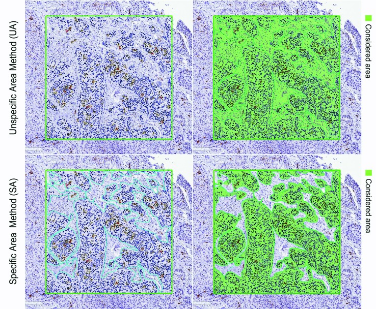
Twenty-six squamous cell carcinomas HIV-positive cases affecting the oropharynx, lips and oral cavity were selected. Immunohistochemistry for CD1a, CD83, and CD207 was performed. The immunohistochemical stains were evaluated by automated examination using a positive pixel count algorithm. A conventional quantification method (unspecific area method; UA) and a novel method (specific area method; SA) were performed obtaining the corresponding density of positive dendritic cells for the intratumoral and peritumoral regions. The Mann-Whitney U test was used to verify the influence of the quantification methods on the positive cell counting according to the evaluated regions. Data were subjected to the ANOVA and Student's t-test to verify the influence of the tumour location, stage, histological grade, and amount of inflammation on the dendritic cells density counting.
The cell quantification method affected the dendritic cells counting independently of the evaluated region (P-value <0.05). Significant differences between methods were also observed according to the tumour features evaluations.
The positive cell quantification method influences the dendritic cells density results. Unlike the conventional method (UA method), the novel SA method avoids non-target areas included in the hotspots improving the reliability and reproducibility of the density cell quantification.
尽管新的数字病理学工具提高了阳性细胞的定量,但文献中定量方法存在异质性。本研究旨在评估并提出一种新的用于鳞状细胞癌的树突状细胞定量方法,并与传统的定量方法进行比较。
选择 26 例 HIV 阳性影响口咽、嘴唇和口腔的鳞状细胞癌病例。进行 CD1a、CD83 和 CD207 的免疫组织化学染色。使用阳性像素计数算法对免疫组化染色进行自动检查评估。进行了传统的定量方法(非特异性区域方法;UA)和新的方法(特异性区域方法;SA),获得肿瘤内和肿瘤周围区域阳性树突状细胞的相应密度。采用曼-惠特尼 U 检验验证定量方法对不同评估区域阳性细胞计数的影响。采用方差分析和学生 t 检验对肿瘤位置、分期、组织学分级和炎症程度对树突状细胞密度计数的影响进行数据验证。
细胞定量方法独立于评估区域影响树突状细胞计数(P 值<0.05)。根据肿瘤特征评估,还观察到方法之间存在显著差异。
阳性细胞定量方法会影响树突状细胞密度结果。与传统方法(UA 方法)不同,新的 SA 方法避免了包括热点中的非目标区域,提高了密度细胞定量的可靠性和可重复性。