Department of Oral and Maxillofacial Pathobiology, Graduate School of Biomedical and Health Sciences, Hiroshima University, Hiroshima, Japan.
Department of Advanced Prosthodontics, Graduate School of Biomedical and Health Sciences, Hiroshima University, Hiroshima, Japan.
Sci Rep. 2020 Mar 5;10(1):4134. doi: 10.1038/s41598-020-60904-8.
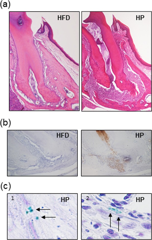
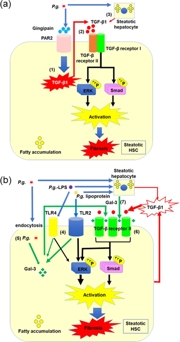
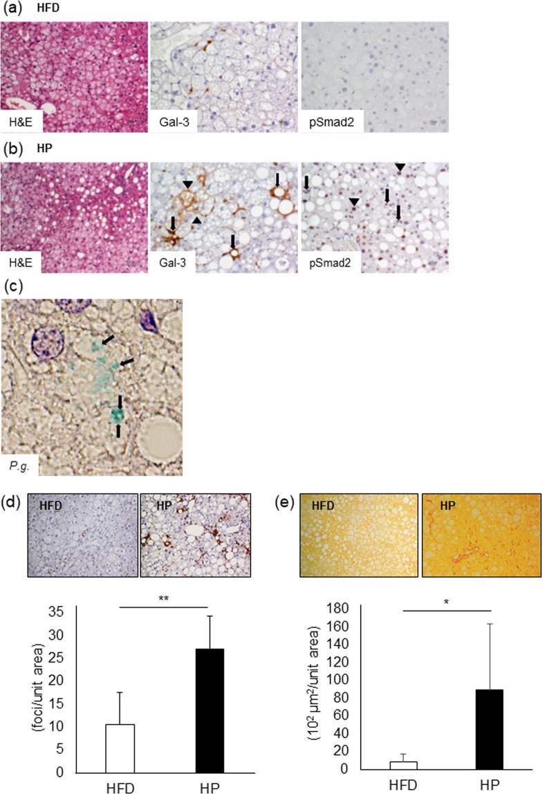
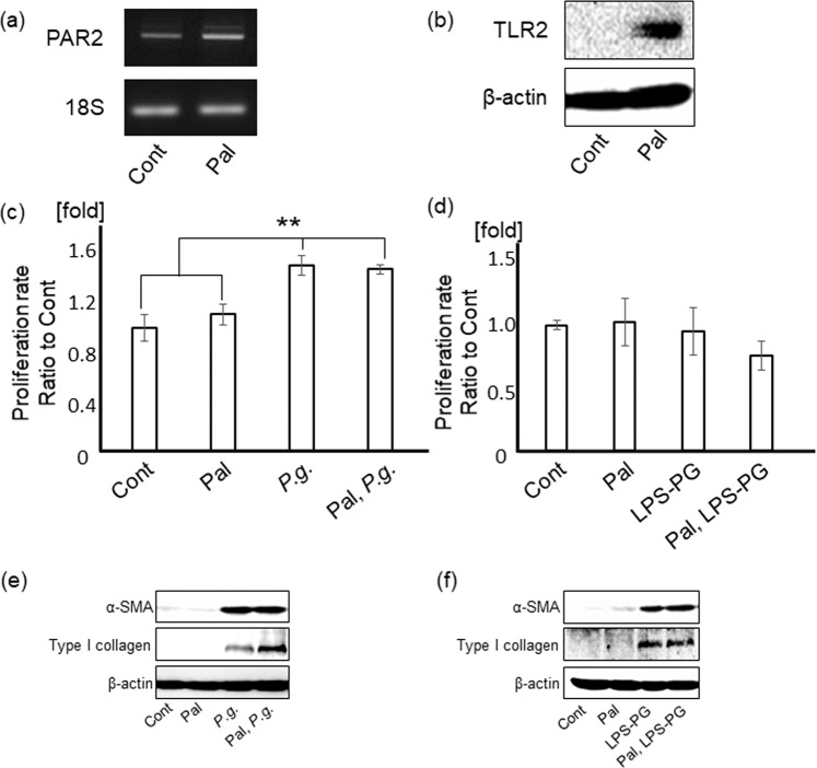
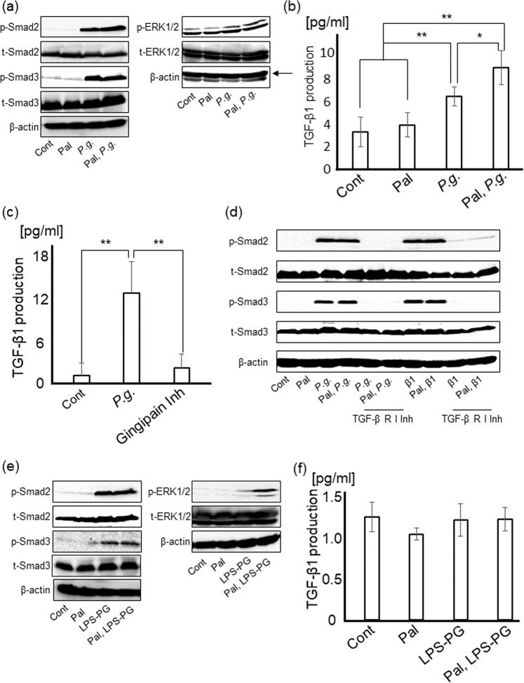
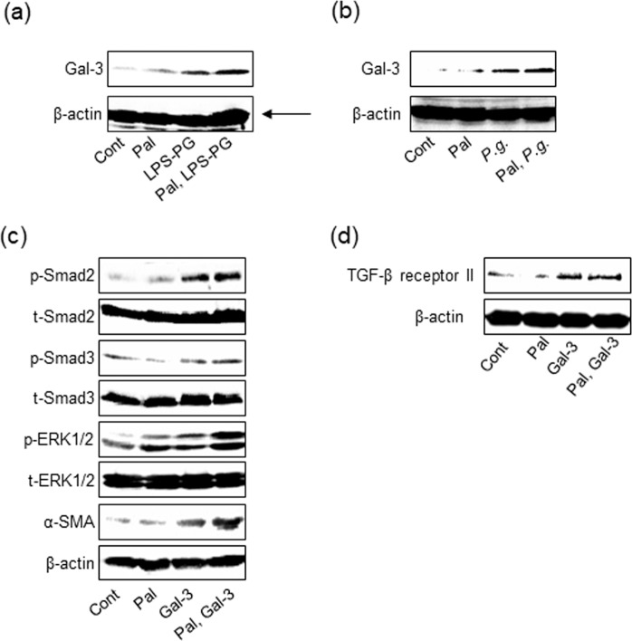
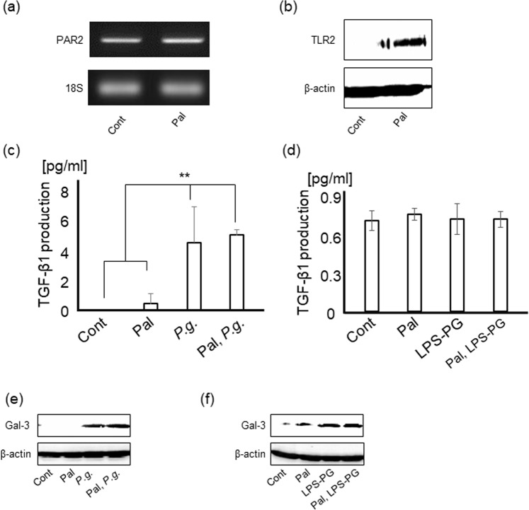
Odontogenic infection of Porphyromonas gingivalis (P.g.), a major periodontal pathogen, exacerbates pathological progression of non-alcoholic steatohepatitis (NASH). In this study, we aimed to clarify the detailed mechanism in which P.g. induced hepatic stellate cells (HSCs; key effector cells in liver fibrosis) activation. In the liver of high fat diet-induced NASH mouse model with P.g. odontogenic infection, immunolocalization of P.g. was detected. The number of hepatic crown-like structure, which was macrophage aggregation and related to liver fibrosis, was drastically increased and fibrosis area was also increased through upregulating immunoexpression of Phosphorylated Smad2 (key signaling molecule of TGF-β1) and Galectin-3. P.g.-secreted trypsin-like enzyme [gingipain; an activator of protease-activated receptor 2 (PAR2)] stimulated HSC proliferation and differentiation through Smad and ERK signaling induced by TGF-β1 produced from HSCs with P.g.-infection. Further, Galectin-3 produced from HSCs with P.g. infection and P.g.-derived LPS/lipoprotein stimulation stabilized TGFβ-receptor II resulting in increasing sensitivity for TGF-β1, finally leading to HSC differentiation via activating Smad and ERK signaling. In addition to them, hepatocytes (main component cells of liver) contributed to HSC activation through TGF-β1 and Galectin-3 production in paracrine manner. Collectively, P.g.-odontogenic infection exacerbates fibrosis of NASH by HSC activation through TGF-β1 and Gal-3 production from HSCs and hepatocytes.
牙龈卟啉单胞菌(P.g.)是一种主要的牙周病原体,其导致的牙源性感染会加剧非酒精性脂肪性肝炎(NASH)的病理进展。在本研究中,我们旨在阐明 P.g. 诱导肝星状细胞(HSCs;肝纤维化的关键效应细胞)活化的详细机制。在高脂肪饮食诱导的 NASH 小鼠模型中,我们检测到 P.g. 牙源性感染部位的免疫定位。通过上调磷酸化 Smad2(TGF-β1 的关键信号分子)和半乳糖凝集素-3 的免疫表达,肝内冠状结构(巨噬细胞聚集与肝纤维化相关)的数量显著增加,纤维化面积也增加。P.g. 分泌的胰蛋白酶样酶(牙龈蛋白酶;蛋白酶激活受体 2(PAR2)的激活剂)通过 HSCs 产生的 TGF-β1 诱导的 Smad 和 ERK 信号刺激 HSC 增殖和分化。此外,受 P.g. 感染的 HSCs 产生的半乳糖凝集素-3 和 P.g. 衍生的 LPS/脂蛋白刺激稳定了 TGFβ-受体 II,从而增加了对 TGF-β1 的敏感性,最终通过激活 Smad 和 ERK 信号导致 HSC 分化。除此之外,肝细胞(肝脏的主要组成细胞)通过旁分泌方式产生 TGF-β1 和半乳糖凝集素-3 促进 HSC 活化。总之,P.g. 牙源性感染通过 HSCs 和肝细胞产生的 TGF-β1 和 Gal-3 促进 HSC 活化,从而加剧 NASH 的纤维化。