Sharma Neekun, Sun Zhe, Hill Michael A, Hans Chetan P
Division of Cardiovascular Medicine, University of Missouri; Dalton Cardiovascular Research Center, University of Missouri.
Medical Pharmacology and Physiology, University of Missouri; Dalton Cardiovascular Research Center, University of Missouri.
J Vis Exp. 2020 Feb 23(156). doi: 10.3791/60515.
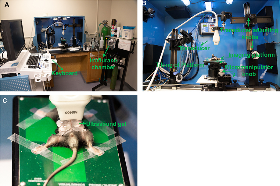
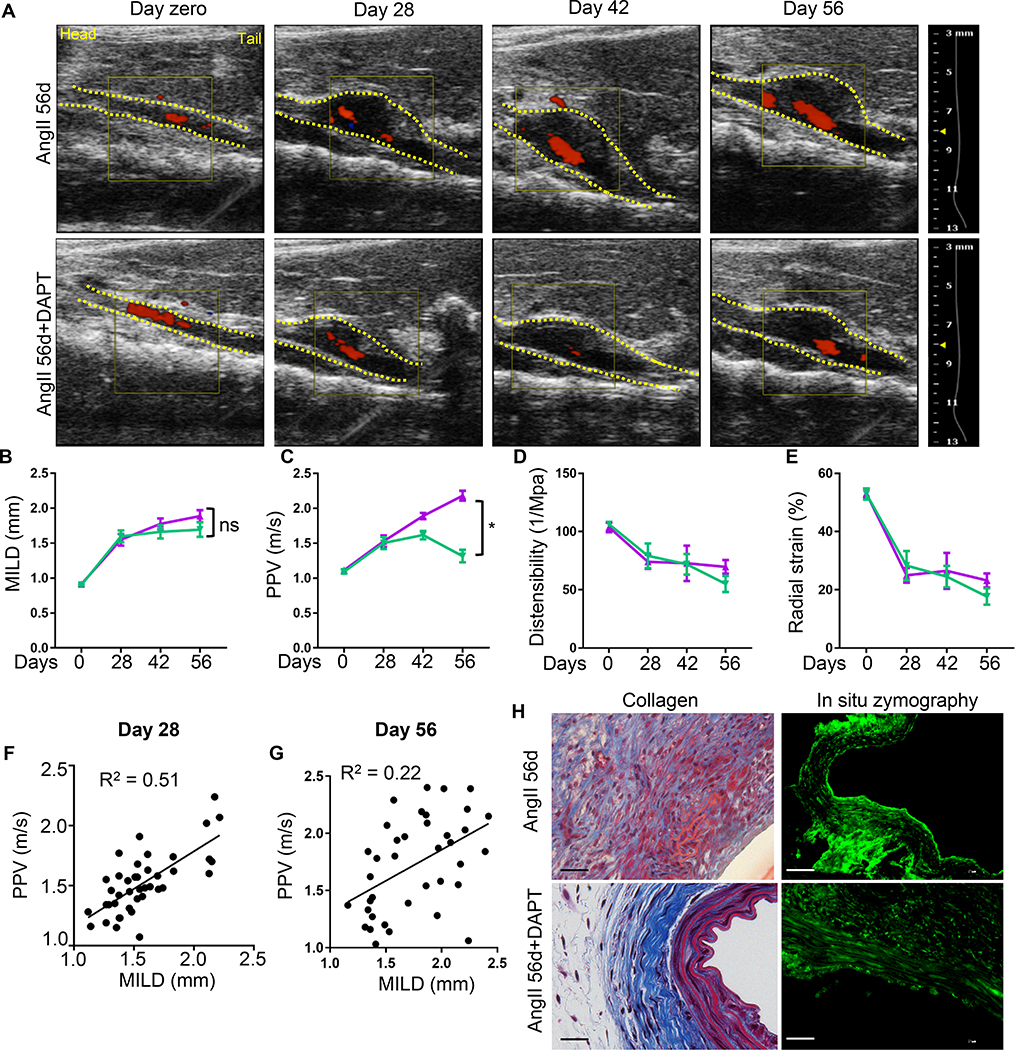
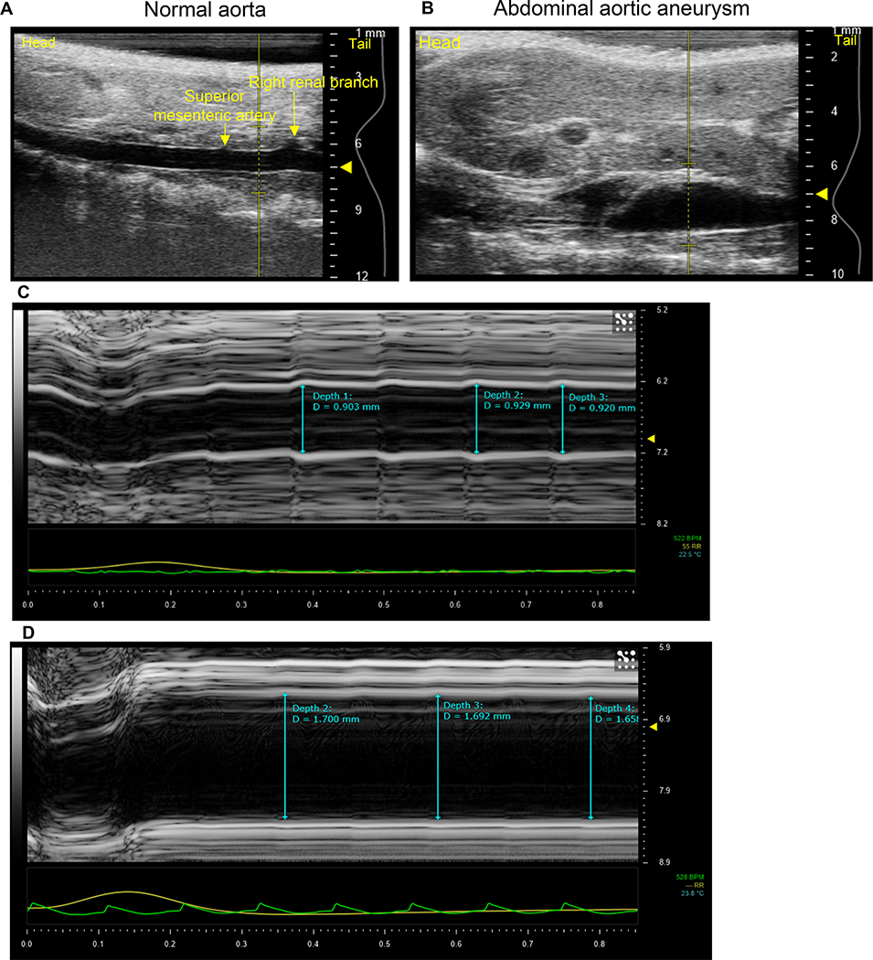
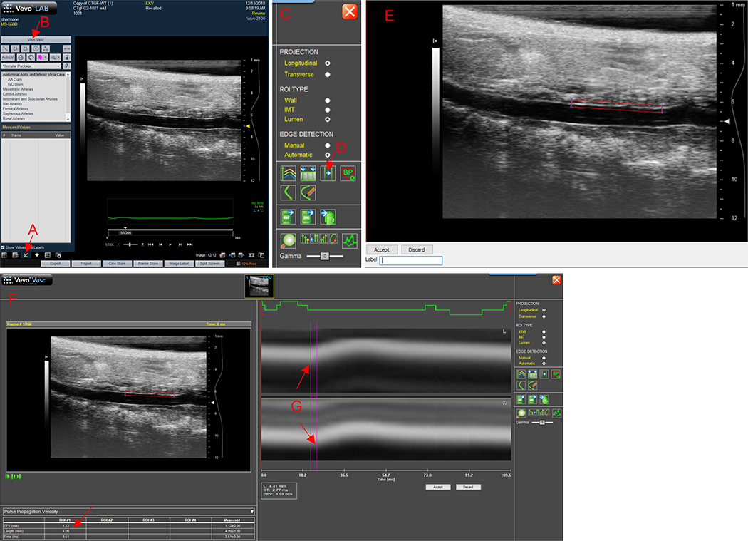
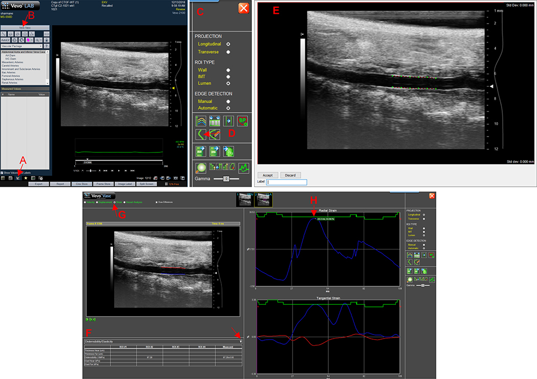
An abdominal aortic aneurysm (AAA) is defined as a localized dilation of the abdominal aorta that exceeds the maximal intraluminal diameter (MILD) by 1.5 times of its original size. Clinical and experimental studies have shown that small aneurysms may rupture, while a subpopulation of large aneurysms may remain stable. Thus, in addition to the measurement of intraluminal diameter of the aorta, knowledge of structural traits of the vessel wall may provide important information to assess the stability of the AAA. Aortic stiffening has recently emerged as a reliable tool to determine early changes in the vascular wall. Pulse propagation velocity (PPV) along with the distensibility and radial strain are highly useful ultrasound-based methods relevant for assessing aortic stiffness. The primary purpose of this protocol is to provide a comprehensive technique for the use of ultrasound imaging system to acquire images and analyze the structural and functional properties of the aorta as determined by MILD, PPV, distensibility and radial strain.
腹主动脉瘤(AAA)被定义为腹主动脉的局限性扩张,其管腔内最大直径(MILD)超过原始大小的1.5倍。临床和实验研究表明,小动脉瘤可能破裂,而一部分大动脉瘤可能保持稳定。因此,除了测量主动脉的管腔内直径外,了解血管壁的结构特征可能为评估腹主动脉瘤的稳定性提供重要信息。主动脉硬化最近已成为确定血管壁早期变化的可靠工具。脉搏传播速度(PPV)以及扩张性和径向应变是与评估主动脉硬度相关的非常有用的基于超声的方法。本方案的主要目的是提供一种综合技术,用于使用超声成像系统获取图像,并分析由MILD、PPV、扩张性和径向应变所确定的主动脉的结构和功能特性。