Department of Psychological and Brain Sciences, Indiana University, Bloomington, Indiana.
Cognitive Science Program, Indiana University, Bloomington, Indiana.
Hum Brain Mapp. 2020 Jun 15;41(9):2249-2262. doi: 10.1002/hbm.24943. Epub 2020 Mar 9.
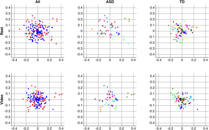
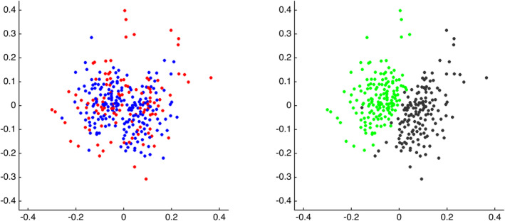
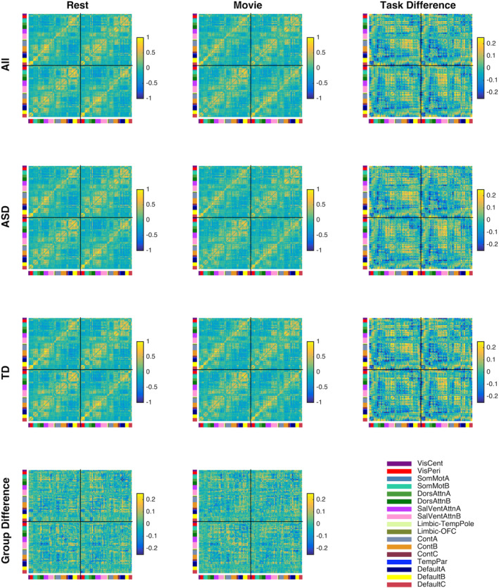
Despite enthusiasm about the potential for using fMRI-based functional connectomes in the development of biomarkers for autism spectrum disorder (ASD), the literature is full of negative findings-failures to distinguish ASD functional connectomes from those of typically developing controls (TD)-and positive findings that are inconsistent across studies. Here, we report on a new study designed to either better differentiate ASD from TD functional connectomes-or, alternatively, to refine our understanding of the factors underlying the current state of affairs. We scanned individuals with ASD and controls both at rest and while watching videos with social content. Using multiband fMRI across repeat sessions, we improved both data quantity and scanning duration by collecting up to 2 hr of data per individual. This is about 50 times the typical number of temporal samples per individual in ASD fcMRI studies. We obtained functional connectomes that were discriminable, allowing for near-perfect individual identification regardless of diagnosis, and equally reliable in both groups. However, contrary to what one might expect, we did not consistently or robustly observe in the ASD group either reductions in similarity to TD functional connectivity (FC) patterns or shared atypical FC patterns. Accordingly, FC-based predictions of diagnosis group achieved accuracy levels around chance. However, using the same approaches to predict scan type (rest vs. video) achieved near-perfect accuracy. Our findings suggest that neither the limitations of resting state as a "task," data resolution, data quantity, or scan duration can be considered solely responsible for failures to differentiate ASD from TD functional connectomes.
尽管人们对使用基于 fMRI 的功能连接组学在自闭症谱系障碍 (ASD) 的生物标志物开发中的应用潜力充满热情,但文献中充满了否定的发现——未能将 ASD 的功能连接组与正常发育对照组 (TD) 的功能连接组区分开来——以及阳性发现,这些发现在不同的研究中并不一致。在这里,我们报告了一项新的研究,旨在更好地区分 ASD 与 TD 的功能连接组,或者,进一步了解当前情况的基础因素。我们对 ASD 患者和对照组个体进行了静息态和观看带有社交内容的视频时的扫描。通过在重复的扫描中使用多频带 fMRI,我们通过每个个体收集多达 2 小时的数据,提高了数据数量和扫描时间。这大约是 ASD fcMRI 研究中每个个体的典型时间样本数量的 50 倍。我们获得了可区分的功能连接组,无论诊断如何,都可以近乎完美地进行个体识别,而且在两组中都同样可靠。然而,与人们的预期相反,我们在 ASD 组中并没有一致或稳健地观察到与 TD 功能连接 (FC) 模式的相似性降低或共享的非典型 FC 模式。因此,基于 FC 的诊断组预测达到了随机水平的准确率。然而,使用相同的方法来预测扫描类型(静息与视频),则达到了近乎完美的准确率。我们的研究结果表明,无论是静息状态作为“任务”的局限性、数据分辨率、数据量还是扫描时间,都不能被认为是唯一导致无法区分 ASD 与 TD 功能连接组的原因。