Cellular and Molecular Research Center, Ahvaz Jundishapur University of Medical Sciences, Ahvaz, Iran.
Department of Anatomical Sciences, Faculty of Medicine, Ahvaz Jundishapur University of Medical Sciences, Ahvaz, Iran.
JBRA Assist Reprod. 2020 Jul 14;24(3):250-256. doi: 10.5935/1518-0557.20190079.
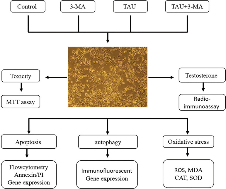
This study evaluated taurine (TAU) effects on autophagy, apoptosis and oxidative stress in mice Leydig TM3 cells.
We treated TM3 cells with TAU (100 µg/mL) or 3-Methyladenine (3-MA, an autophagy inhibitor) for 24 h, and assessed cell viability, testosterone level, oxidative stress, apoptosis, and autophagy.
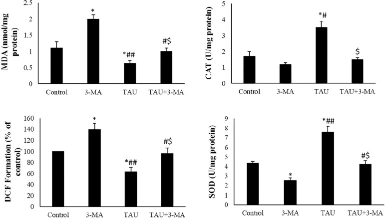
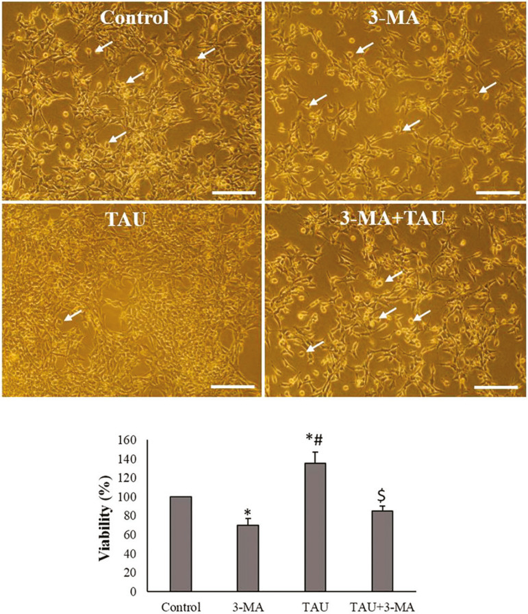
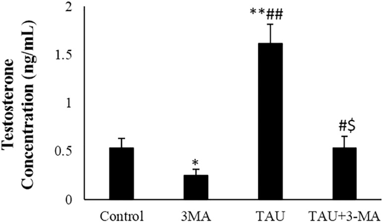
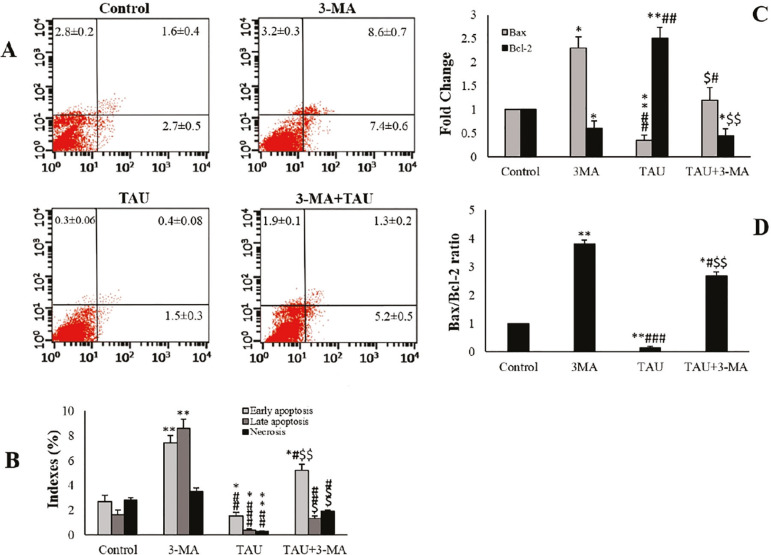
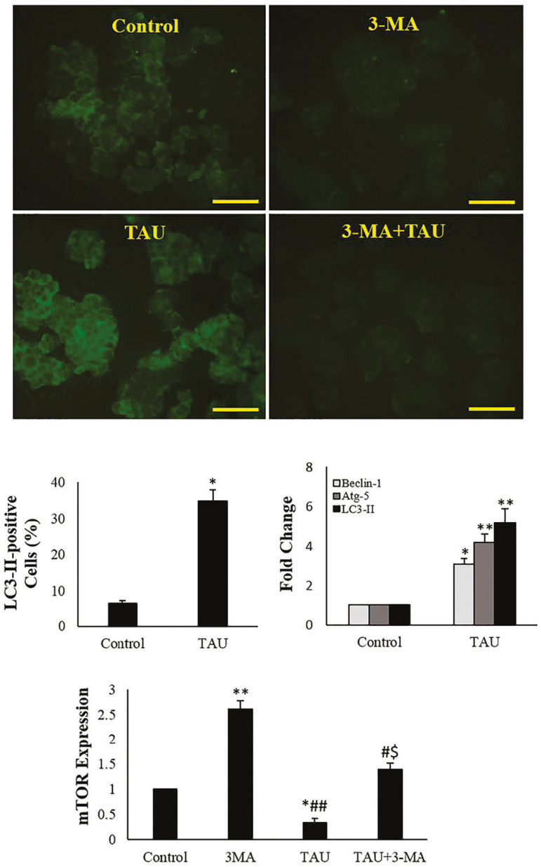
The results showed that TAU markedly increased cell viability, testosterone levels, expression of autophagy-related genes and percentage of LC3-II-positive cells. TAU significantly reduced malondialdehyde (MDA) contents and reactive oxygen species (ROS) levels and increased the activities of SOD (superoxide dismutase) and CAT (Catalase) enzymes in the TM3 cells. TAU in the presence of autophagy inhibitor (3-MA) increased oxidative stress and decreased testosterone levels.
The results showed that autophagy might be involved in TAU-increased testosterone levels in mice Leydig TM3 cells.
本研究评估牛磺酸(TAU)对小鼠 LeydigTM3 细胞自噬、凋亡和氧化应激的影响。
我们用 TAU(100μg/ml)或 3-甲基腺嘌呤(3-MA,自噬抑制剂)处理 TM3 细胞 24 小时,评估细胞活力、睾酮水平、氧化应激、凋亡和自噬。
结果表明,TAU 显著增加细胞活力、睾酮水平、自噬相关基因的表达和 LC3-II 阳性细胞的百分比。TAU 显著降低丙二醛(MDA)含量和活性氧(ROS)水平,并增加 TM3 细胞中超氧化物歧化酶(SOD)和过氧化氢酶(CAT)的活性。在自噬抑制剂(3-MA)存在的情况下,TAU 增加了氧化应激并降低了睾酮水平。
结果表明,自噬可能参与了 TAU 增加小鼠 LeydigTM3 细胞睾酮水平的过程。