Institute for Ophthalmic Research, University of Tübingen, Tübingen, Germany.
Centre for Integrative Neuroscience (CIN), University of Tübingen, Tübingen, Germany.
Sci Rep. 2020 Mar 10;10(1):4399. doi: 10.1038/s41598-020-60214-z.
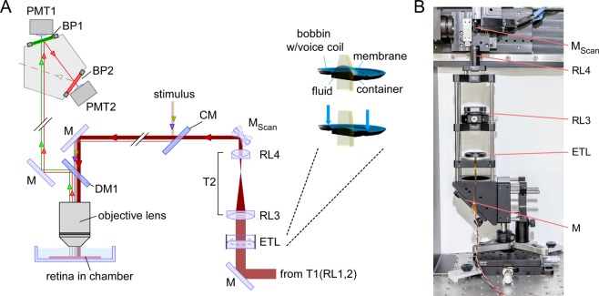
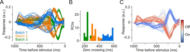
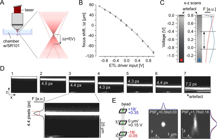
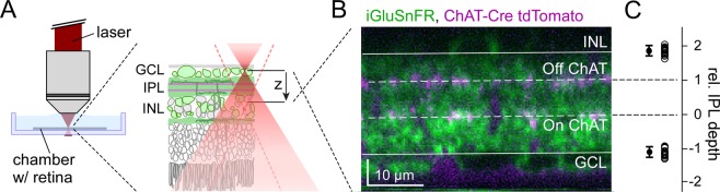
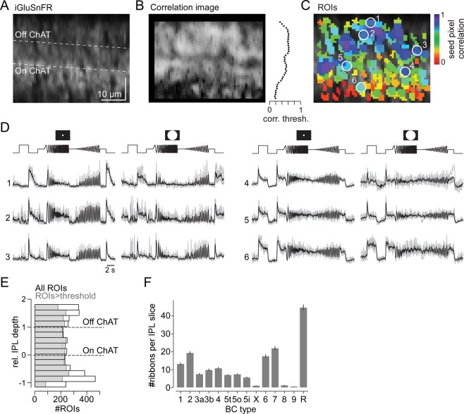
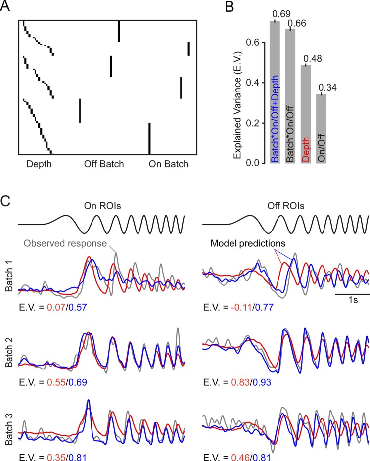
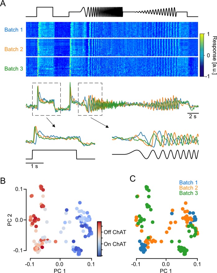
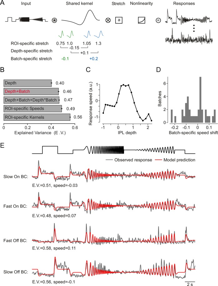
The retina decomposes visual stimuli into parallel channels that encode different features of the visual environment. Central to this computation is the synaptic processing in a dense layer of neuropil, the so-called inner plexiform layer (IPL). Here, different types of bipolar cells stratifying at distinct depths relay the excitatory feedforward drive from photoreceptors to amacrine and ganglion cells. Current experimental techniques for studying processing in the IPL do not allow imaging the entire IPL simultaneously in the intact tissue. Here, we extend a two-photon microscope with an electrically tunable lens allowing us to obtain optical vertical slices of the IPL, which provide a complete picture of the response diversity of bipolar cells at a "single glance". The nature of these axial recordings additionally allowed us to isolate and investigate batch effects, i.e. inter-experimental variations resulting in systematic differences in response speed. As a proof of principle, we developed a simple model that disentangles biological from experimental causes of variability and allowed us to recover the characteristic gradient of response speeds across the IPL with higher precision than before. Our new framework will make it possible to study the computations performed in the central synaptic layer of the retina more efficiently.
视网膜将视觉刺激分解为平行的通道,这些通道编码视觉环境的不同特征。这种计算的核心是在所谓的内丛状层(IPL)中神经突起的密集层中的突触处理。在这里,分层在不同深度的不同类型的双极细胞将光感受器的兴奋性前馈驱动中继到无长突细胞和神经节细胞。目前用于研究 IPL 中处理的实验技术不允许在完整组织中同时对整个 IPL 进行成像。在这里,我们扩展了具有电可调透镜的双光子显微镜,使我们能够获得 IPL 的光学垂直切片,这些切片提供了双极细胞反应多样性的完整图片,只需“一瞥”即可。这些轴向记录的性质还允许我们分离和研究批处理效应,即由于响应速度的系统差异而导致的实验间变化。作为原理验证,我们开发了一个简单的模型,该模型将生物和实验引起的可变性分开,并使我们能够比以前更高的精度恢复 IPL 中响应速度的特征梯度。我们的新框架将使研究视网膜中央突触层中执行的计算更加高效。