• 文献检索
  • 文档翻译
  • 深度研究
  • 学术资讯
  • Suppr Zotero 插件Zotero 插件
  • 邀请有礼
  • 套餐&价格
  • 历史记录
应用&插件
Suppr Zotero 插件Zotero 插件浏览器插件Mac 客户端Windows 客户端微信小程序
定价
高级版会员购买积分包购买API积分包
服务
文献检索文档翻译深度研究API 文档MCP 服务
关于我们
关于 Suppr公司介绍联系我们用户协议隐私条款
关注我们

Suppr 超能文献

核心技术专利:CN118964589B侵权必究
粤ICP备2023148730 号-1Suppr @ 2026

文献检索

告别复杂PubMed语法,用中文像聊天一样搜索,搜遍4000万医学文献。AI智能推荐,让科研检索更轻松。

立即免费搜索

文件翻译

保留排版,准确专业,支持PDF/Word/PPT等文件格式,支持 12+语言互译。

免费翻译文档

深度研究

AI帮你快速写综述,25分钟生成高质量综述,智能提取关键信息,辅助科研写作。

立即免费体验

糖尿病状况及AGE/RAGE信号传导对心脏成纤维细胞迁移的影响

The Impact of Diabetic Conditions and AGE/RAGE Signaling on Cardiac Fibroblast Migration.

作者信息

Burr Stephanie D, Harmon Mallory B, Jr James A Stewart

机构信息

Department of BioMolecular Sciences, School of Pharmacy, The University of Mississippi, Oxford, MS, United States.

出版信息

Front Cell Dev Biol. 2020 Feb 25;8:112. doi: 10.3389/fcell.2020.00112. eCollection 2020.

DOI:10.3389/fcell.2020.00112
PMID:32158758
原文链接:https://pmc.ncbi.nlm.nih.gov/articles/PMC7052116/
Abstract

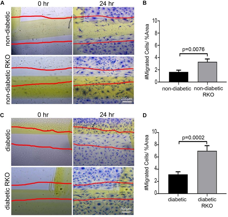
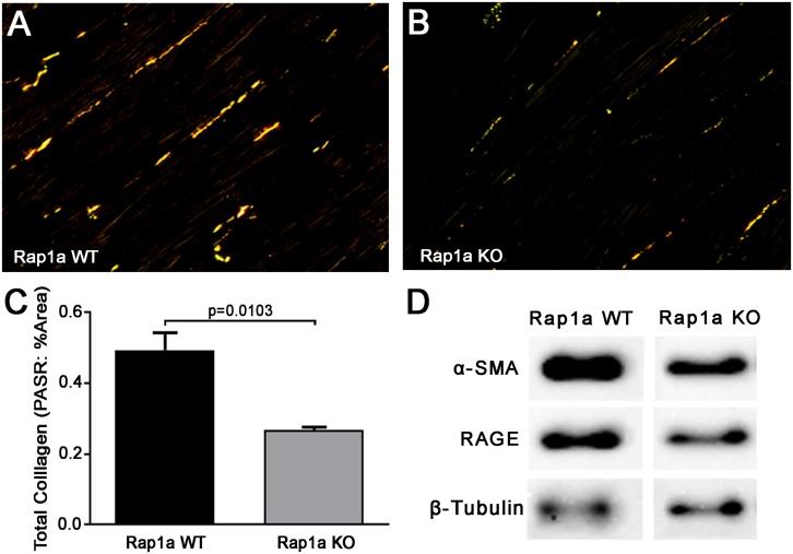
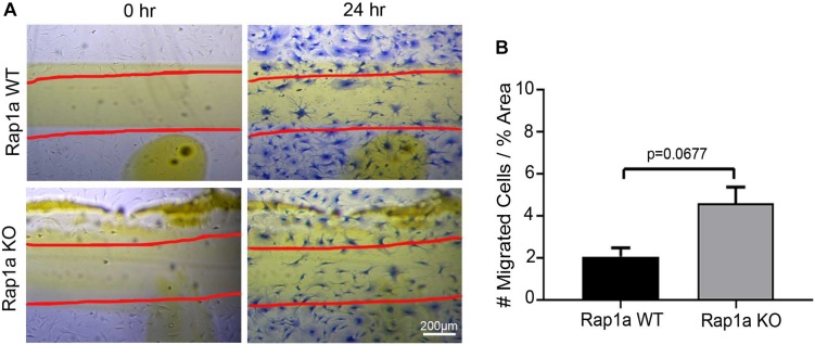
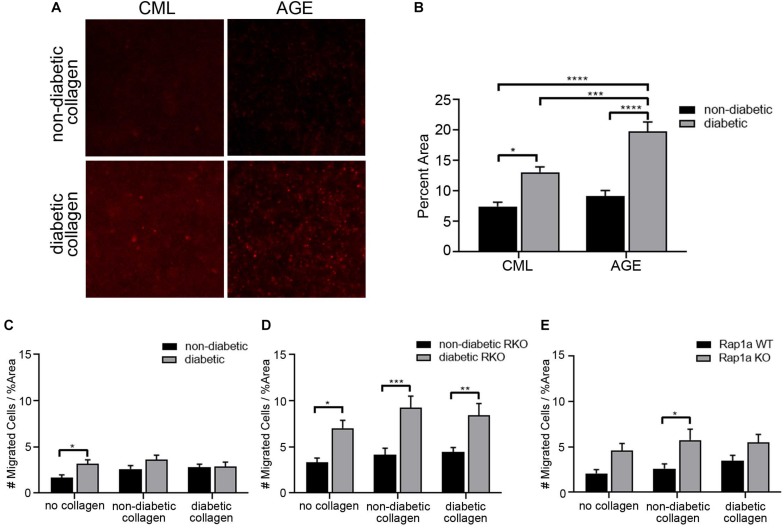
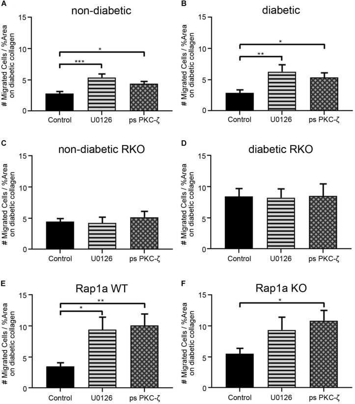
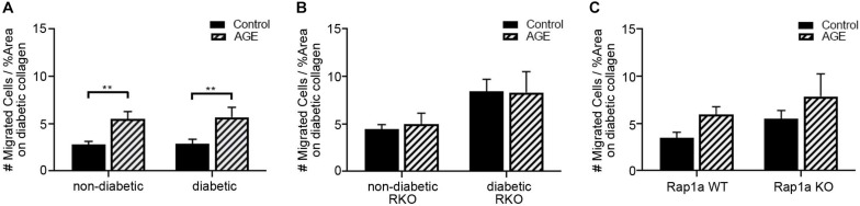
Diabetic individuals have an increased risk for developing cardiovascular disease due to stiffening of the left ventricle (LV), which is thought to occur, in part, by increased AGE/RAGE signaling inducing fibroblast differentiation. Advanced glycated end-products (AGEs) accumulate within the body over time, and under hyperglycemic conditions, the formation and accumulation of AGEs is accelerated. AGEs exert their effect by binding to their receptor (RAGE) and can induce myofibroblast differentiation, leading to increased cell migration. Previous studies have focused on fibroblast migration during wound healing, in which diabetics have impaired fibroblast migration compared to healthy individuals. However, the impact of diabetic conditions as well as AGE/RAGE signaling has not been extensively studied in cardiac fibroblasts. Therefore, the goal of this study was to determine how the AGE/RAGE signaling pathway impacts cell migration in non-diabetic and diabetic cardiac fibroblasts. Cardiac fibroblasts were isolated from non-diabetic and diabetic mice with and without functional RAGE and used to perform a migration assay. Cardiac fibroblasts were plated on plastic, non-diabetic, or diabetic collagen, and when confluency was reached, a line of migration was generated by scratching the plate and followed by treatment with pharmacological agents that modify AGE/RAGE signaling. Modification of the AGE/RAGE signaling cascade was done with ERK1/2 and PKC-ζ inhibitors as well as treatment with exogenous AGEs. Diabetic fibroblasts displayed an increase in migration compared to non-diabetic fibroblasts whereas inhibiting the AGE/RAGE signaling pathway resulted in a significant increase in migration. The results indicate that the AGE/RAGE signaling cascade causes a decrease in cardiac fibroblast migration and altering the pathway will produce alterations in cardiac fibroblast migration.

摘要

糖尿病个体因左心室(LV)僵硬而患心血管疾病的风险增加,这种僵硬被认为部分是由于AGE/RAGE信号增加诱导成纤维细胞分化所致。晚期糖基化终产物(AGEs)会随着时间在体内积累,在高血糖条件下,AGEs的形成和积累会加速。AGEs通过与其受体(RAGE)结合发挥作用,并可诱导肌成纤维细胞分化,导致细胞迁移增加。先前的研究集中在伤口愈合过程中的成纤维细胞迁移,与健康个体相比,糖尿病患者的成纤维细胞迁移受损。然而,糖尿病状态以及AGE/RAGE信号的影响在心脏成纤维细胞中尚未得到广泛研究。因此,本研究的目的是确定AGE/RAGE信号通路如何影响非糖尿病和糖尿病心脏成纤维细胞的细胞迁移。从具有和不具有功能性RAGE的非糖尿病和糖尿病小鼠中分离出心脏成纤维细胞,并用于进行迁移试验。将心脏成纤维细胞接种在塑料、非糖尿病或糖尿病胶原蛋白上,当达到汇合时,通过刮擦平板产生一条迁移线,然后用改变AGE/RAGE信号的药物进行处理。使用ERK1/2和PKC-ζ抑制剂以及外源性AGEs处理来改变AGE/RAGE信号级联。与非糖尿病成纤维细胞相比,糖尿病成纤维细胞的迁移增加,而抑制AGE/RAGE信号通路导致迁移显著增加。结果表明,AGE/RAGE信号级联导致心脏成纤维细胞迁移减少,改变该通路将导致心脏成纤维细胞迁移发生改变。

https://cdn.ncbi.nlm.nih.gov/pmc/blobs/baba/7052116/8b8ae5331c14/fcell-08-00112-g007.jpg
https://cdn.ncbi.nlm.nih.gov/pmc/blobs/baba/7052116/bd22a956cd4f/fcell-08-00112-g001.jpg
https://cdn.ncbi.nlm.nih.gov/pmc/blobs/baba/7052116/0a06f07687c4/fcell-08-00112-g002.jpg
https://cdn.ncbi.nlm.nih.gov/pmc/blobs/baba/7052116/ea4b5a665170/fcell-08-00112-g003.jpg
https://cdn.ncbi.nlm.nih.gov/pmc/blobs/baba/7052116/9d61441875c3/fcell-08-00112-g004.jpg
https://cdn.ncbi.nlm.nih.gov/pmc/blobs/baba/7052116/d3b7cf831d4a/fcell-08-00112-g005.jpg
https://cdn.ncbi.nlm.nih.gov/pmc/blobs/baba/7052116/dd2ab62dff90/fcell-08-00112-g006.jpg
https://cdn.ncbi.nlm.nih.gov/pmc/blobs/baba/7052116/8b8ae5331c14/fcell-08-00112-g007.jpg

相似文献

1
The Impact of Diabetic Conditions and AGE/RAGE Signaling on Cardiac Fibroblast Migration.糖尿病状况及AGE/RAGE信号传导对心脏成纤维细胞迁移的影响
Front Cell Dev Biol. 2020 Feb 25;8:112. doi: 10.3389/fcell.2020.00112. eCollection 2020.
2
Rap1a Overlaps the AGE/RAGE Signaling Cascade to Alter Expression of α-SMA, p-NF-κB, and p-PKC-ζ in Cardiac Fibroblasts Isolated from Type 2 Diabetic Mice.Rap1a 与 AGE/RAGE 信号级联重叠,改变 2 型糖尿病小鼠心脏成纤维细胞中 α-SMA、p-NF-κB 和 p-PKC-ζ 的表达。
Cells. 2021 Mar 4;10(3):557. doi: 10.3390/cells10030557.
3
Rap1a Regulates Cardiac Fibroblast Contraction of 3D Diabetic Collagen Matrices by Increased Activation of the AGE/RAGE Cascade.Rap1a 通过增加 AGE/RAGE 级联反应的激活来调节 3D 糖尿病胶原基质中心律失常成纤维细胞的收缩。
Cells. 2021 May 22;10(6):1286. doi: 10.3390/cells10061286.
4
Extracellular matrix components isolated from diabetic mice alter cardiac fibroblast function through the AGE/RAGE signaling cascade.从糖尿病小鼠中分离出的细胞外基质成分通过 AGE/RAGE 信号级联改变心肌成纤维细胞的功能。
Life Sci. 2020 Jun 1;250:117569. doi: 10.1016/j.lfs.2020.117569. Epub 2020 Mar 19.
5
Rap1a Activity Elevated the Impact of Endogenous AGEs in Diabetic Collagen to Stimulate Increased Myofibroblast Transition and Oxidative Stress.Rap1a 活性升高了糖尿病胶原中内源性 AGEs 的影响,从而刺激肌成纤维细胞过渡和氧化应激增加。
Int J Mol Sci. 2022 Apr 19;23(9):4480. doi: 10.3390/ijms23094480.
6
Molecular mechanisms of AGE/RAGE-mediated fibrosis in the diabetic heart.糖尿病心脏中晚期糖基化终末产物/晚期糖基化终末产物受体介导的纤维化的分子机制
World J Diabetes. 2014 Dec 15;5(6):860-7. doi: 10.4239/wjd.v5.i6.860.
7
Regenerative and protective effects of dMSC-sEVs on high-glucose-induced senescent fibroblasts by suppressing RAGE pathway and activating Smad pathway.人骨髓间充质干细胞来源的外泌体通过抑制 RAGE 通路和激活 Smad 通路对高糖诱导的衰老成纤维细胞的再生和保护作用。
Stem Cell Res Ther. 2020 Apr 29;11(1):166. doi: 10.1186/s13287-020-01681-z.
8
Atorvastatin prevents advanced glycation end products (AGEs)-induced cardiac fibrosis via activating peroxisome proliferator-activated receptor gamma (PPAR-γ).阿托伐他汀通过激活过氧化物酶体增殖物激活受体γ(PPAR-γ)来预防晚期糖基化终产物(AGEs)诱导的心脏纤维化。
Metabolism. 2016 Apr;65(4):441-53. doi: 10.1016/j.metabol.2015.11.007. Epub 2015 Nov 27.
9
RAGE and TGF-β1 Cross-Talk Regulate Extracellular Matrix Turnover and Cytokine Synthesis in AGEs Exposed Fibroblast Cells.RAGE与TGF-β1的相互作用调节晚期糖基化终末产物作用下成纤维细胞的细胞外基质周转和细胞因子合成。
PLoS One. 2016 Mar 25;11(3):e0152376. doi: 10.1371/journal.pone.0152376. eCollection 2016.
10
Cellular dysfunction in the diabetic fibroblast: impairment in migration, vascular endothelial growth factor production, and response to hypoxia.糖尿病成纤维细胞中的细胞功能障碍:迁移、血管内皮生长因子产生及对缺氧反应的受损。
Am J Pathol. 2003 Jan;162(1):303-12. doi: 10.1016/S0002-9440(10)63821-7.

引用本文的文献

1
The RAGE Inhibitor TTP488 (Azeliragon) Improves Diabetic Bladder Dysfunction in Leptin-Deficient Obese Mice.晚期糖基化终末产物受体抑制剂TTP488(阿泽利拉贡)可改善瘦素缺乏型肥胖小鼠的糖尿病膀胱功能障碍。
Antioxidants (Basel). 2025 Jun 27;14(7):793. doi: 10.3390/antiox14070793.
2
Inhibition of high glucose-induced cardiac fibroblast activation: an effective treatment for diabetic cardiomyopathy using Chinese herbal medicine.抑制高糖诱导的心脏成纤维细胞活化:一种使用中药治疗糖尿病性心肌病的有效方法。
Front Pharmacol. 2025 Jan 27;16:1523014. doi: 10.3389/fphar.2025.1523014. eCollection 2025.
3
Receptor activity-modifying protein 1 regulates the differentiation of mouse skin fibroblasts by downregulating α-SMA expression via suppression of high mobility group AT-hook 1 to promote skin wound repair.

本文引用的文献

1
Increased fractal dimension of left ventricular trabeculations is associated with subclinical diastolic dysfunction in patients with type-2 diabetes mellitus.2型糖尿病患者左心室小梁的分形维数增加与亚临床舒张功能障碍有关。
Int J Cardiovasc Imaging. 2019 Apr;35(4):665-673. doi: 10.1007/s10554-018-1492-0. Epub 2018 Nov 14.
2
Receptor for advanced glycation end products is targeted by FBXO10 for ubiquitination and degradation.晚期糖基化终末产物受体被FBXO10靶向进行泛素化和降解。
FASEB J. 2017 Sep;31(9):3894-3903. doi: 10.1096/fj.201700031R. Epub 2017 May 17.
3
Age-dependent development of left ventricular wall thickness in type 2 diabetic (db/db) mice is associated with elevated low-density lipoprotein and triglyceride serum levels.
受体活性修饰蛋白1通过抑制高迁移率族AT-钩蛋白1下调α-平滑肌肌动蛋白表达,从而调节小鼠皮肤成纤维细胞分化,促进皮肤伤口修复。
Burns Trauma. 2025 Jan 21;13:tkae068. doi: 10.1093/burnst/tkae068. eCollection 2025.
4
Glycation in the cardiomyocyte.心肌细胞中的糖基化作用。
Vitam Horm. 2024;125:47-88. doi: 10.1016/bs.vh.2024.04.005. Epub 2024 May 24.
5
Identification of potential shared gene signatures between gastric cancer and type 2 diabetes: a data-driven analysis.胃癌与2型糖尿病潜在共享基因特征的鉴定:一项数据驱动分析
Front Med (Lausanne). 2024 Jun 6;11:1382004. doi: 10.3389/fmed.2024.1382004. eCollection 2024.
6
[Differential expressions of endoplasmic reticulum stress-associated genes in aortic dissection and their correlation with immune cell infiltration].[内质网应激相关基因在主动脉夹层中的差异表达及其与免疫细胞浸润的相关性]
Nan Fang Yi Ke Da Xue Xue Bao. 2024 May 20;44(5):859-866. doi: 10.12122/j.issn.1673-4254.2024.05.07.
7
Development of raft-forming liquid formulations loaded with ginger extract-solid dispersion for treatment of gastric ulceration.用于治疗胃溃疡的载姜提取物-固体分散体的筏状形成液体制剂的研发。
Heliyon. 2024 May 23;10(11):e31803. doi: 10.1016/j.heliyon.2024.e31803. eCollection 2024 Jun 15.
8
Elucidating the anti-hypertensive mechanisms of Uncaria rhynchophylla-Alisma plantago-aquatica L: an integrated network pharmacology, cluster analysis, and molecular docking approach.阐明钩藤-泽泻的抗高血压机制:综合网络药理学、聚类分析和分子对接方法
Front Chem. 2024 Feb 29;12:1356458. doi: 10.3389/fchem.2024.1356458. eCollection 2024.
9
Central role of cardiac fibroblasts in myocardial fibrosis of diabetic cardiomyopathy.心脏成纤维细胞在糖尿病心肌病心肌纤维化中的核心作用。
Front Endocrinol (Lausanne). 2023 Mar 31;14:1162754. doi: 10.3389/fendo.2023.1162754. eCollection 2023.
10
Mechanism of Datura metel on sinus bradycardia based on network pharmacology and molecular docking.基于网络药理学和分子对接技术研究曼陀罗对窦性心动过缓的作用机制。
Medicine (Baltimore). 2022 Dec 9;101(49):e32190. doi: 10.1097/MD.0000000000032190.
2型糖尿病(db/db)小鼠左心室壁厚度的年龄依赖性发展与血清低密度脂蛋白和甘油三酯水平升高有关。
Heart Vessels. 2017 Aug;32(8):1025-1031. doi: 10.1007/s00380-017-0978-3. Epub 2017 Apr 9.
4
Protective effect of mangiferin on myocardial ischemia-reperfusion injury in streptozotocin-induced diabetic rats: role of AGE-RAGE/MAPK pathways.芒果苷对链脲佐菌素诱导的糖尿病大鼠心肌缺血再灌注损伤的保护作用:AGE-RAGE/MAPK 通路的作用。
Sci Rep. 2017 Feb 9;7:42027. doi: 10.1038/srep42027.
5
Hyperglycemia reduces integrin subunits alpha v and alpha 5 on the surface of dermal fibroblasts contributing to deficient migration.高血糖会降低真皮成纤维细胞表面的整合素亚基αv和α5,导致迁移能力不足。
Mol Cell Biochem. 2016 Oct;421(1-2):19-28. doi: 10.1007/s11010-016-2780-4. Epub 2016 Aug 18.
6
Extracellular Matrix and Dermal Fibroblast Function in the Healing Wound.愈合伤口中的细胞外基质与真皮成纤维细胞功能
Adv Wound Care (New Rochelle). 2016 Mar 1;5(3):119-136. doi: 10.1089/wound.2014.0561.
7
Advanced glycation end products upregulate lysyl oxidase and endothelin-1 in human aortic endothelial cells via parallel activation of ERK1/2-NF-κB and JNK-AP-1 signaling pathways.晚期糖基化终末产物通过ERK1/2-NF-κB和JNK-AP-1信号通路的平行激活上调人主动脉内皮细胞中的赖氨酰氧化酶和内皮素-1。
Cell Mol Life Sci. 2016 Apr;73(8):1685-98. doi: 10.1007/s00018-015-2091-z. Epub 2015 Dec 8.
8
Fibroblast heterogeneity and its implications for engineering organotypic skin models in vitro.成纤维细胞异质性及其对体外器官型皮肤模型工程的影响。
Eur J Cell Biol. 2015 Nov;94(11):483-512. doi: 10.1016/j.ejcb.2015.08.001. Epub 2015 Aug 14.
9
Metalloproteinases and Wound Healing.金属蛋白酶与伤口愈合
Adv Wound Care (New Rochelle). 2015 Apr 1;4(4):225-234. doi: 10.1089/wound.2014.0581.
10
Molecular mechanisms of AGE/RAGE-mediated fibrosis in the diabetic heart.糖尿病心脏中晚期糖基化终末产物/晚期糖基化终末产物受体介导的纤维化的分子机制
World J Diabetes. 2014 Dec 15;5(6):860-7. doi: 10.4239/wjd.v5.i6.860.