Rosser J, Bachmann B, Jordan C, Ribitsch I, Haltmayer E, Gueltekin S, Junttila S, Galik B, Gyenesei A, Haddadi B, Harasek M, Egerbacher M, Ertl P, Jenner F
Faculty of Technical Chemistry, Vienna University of Technology, Getreidemarkt 9, 1060 Vienna, Austria.
Department of Equine Surgery, University of Veterinary Medicine, Veterinärplatz 1, 1210 Vienna, Austria.
Mater Today Bio. 2019 Aug 19;4:100023. doi: 10.1016/j.mtbio.2019.100023. eCollection 2019 Sep.
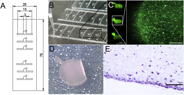
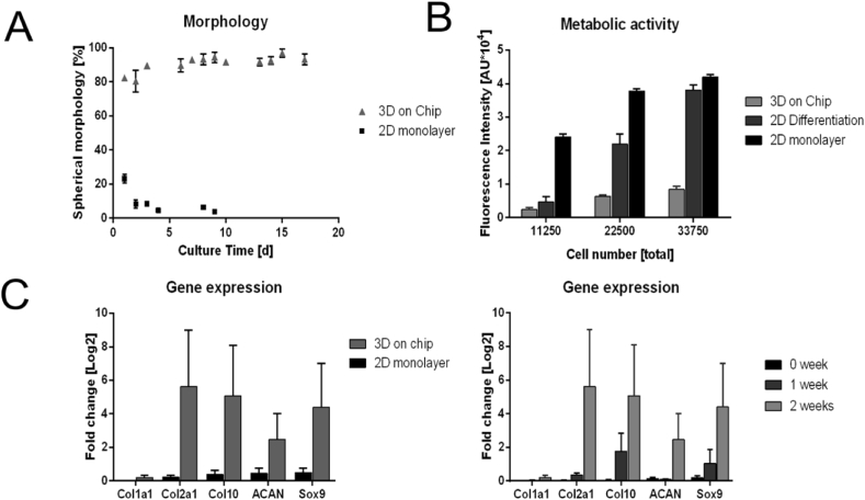
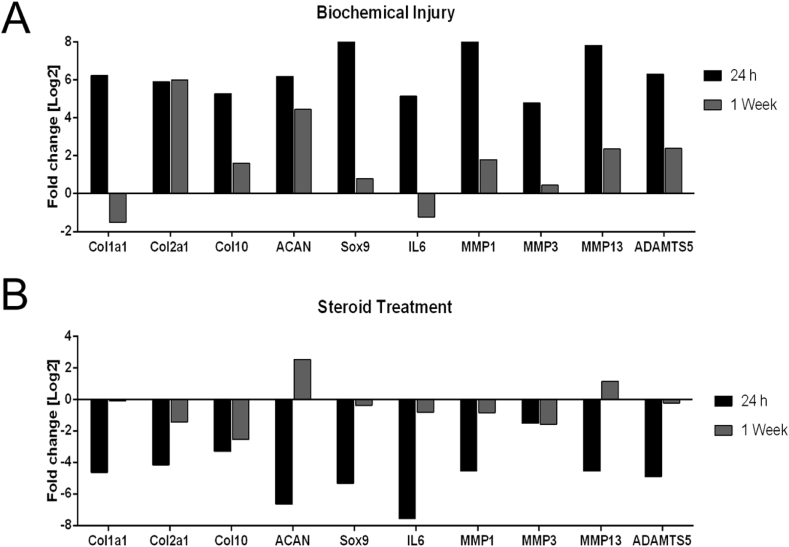
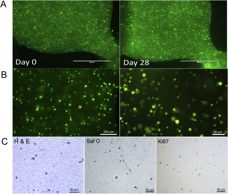
In this work, we describe a microfluidic three-dimensional (3D) chondrocyte culture mimicking articular chondrocyte morphology, cell distribution, metabolism, and gene expression. This has been accomplished by establishing a physiologic nutrient diffusion gradient across the simulated matrix, while geometric design constraints of the microchambers drive native-like cellular behavior. Primary equine chondrocytes remained viable for the extended culture time of 3 weeks and maintained the low metabolic activity and high Sox9, aggrecan, and Col2 expression typical of articular chondrocytes. Our microfluidic 3D chondrocyte microtissues were further exposed to inflammatory cytokines to establish an animal-free, osteoarthritis model. Results of our study indicate that our microtissue model emulates the basic characteristics of native cartilage and responds to biochemical injury, thus providing a new foundation for exploration of osteoarthritis pathophysiology in both human and veterinary patients.
在这项工作中,我们描述了一种微流控三维(3D)软骨细胞培养方法,该方法模拟了关节软骨细胞的形态、细胞分布、代谢和基因表达。这是通过在模拟基质上建立生理营养物质扩散梯度来实现的,而微腔室的几何设计限制驱动了类似天然的细胞行为。原代马软骨细胞在长达3周的延长培养时间内保持存活,并维持了关节软骨细胞典型的低代谢活性以及高Sox9、聚集蛋白聚糖和Col2表达。我们的微流控3D软骨细胞微组织进一步暴露于炎性细胞因子中,以建立一个无动物的骨关节炎模型。我们的研究结果表明,我们的微组织模型模拟了天然软骨的基本特征,并对生化损伤做出反应,从而为探索人类和兽医患者的骨关节炎病理生理学提供了新的基础。