Ranasinghe Kamalini G, Cha Jungho, Iaccarino Leonardo, Hinkley Leighton B, Beagle Alexander J, Pham Julie, Jagust William J, Miller Bruce L, Rankin Katherine P, Rabinovici Gil D, Vossel Keith A, Nagarajan Srikantan S
Memory and Aging Center, Department of Neurology, University of California, San Francisco, San Francisco, CA 94158, USA.
Department Radiology and Biomedical Imaging, University of California, San Francisco, San Francisco, CA 94143, USA.
Sci Transl Med. 2020 Mar 11;12(534). doi: 10.1126/scitranslmed.aaz4069.
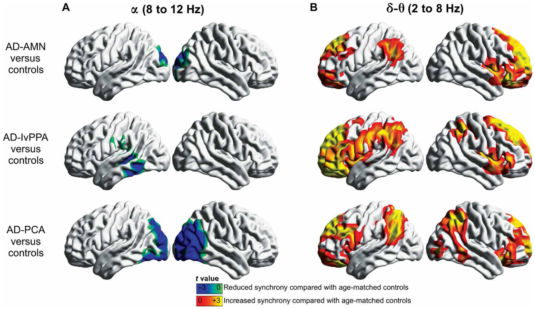
Neural synchrony is intricately balanced in the normal resting brain but becomes altered in Alzheimer's disease (AD). To determine the neurophysiological manifestations associated with molecular biomarkers of AD neuropathology, in patients with AD, we used magnetoencephalographic imaging (MEGI) and positron emission tomography with amyloid-beta (Aβ) and TAU tracers. We found that alpha oscillations (8 to 12 Hz) were hyposynchronous in occipital and posterior temporoparietal cortices, whereas delta-theta oscillations (2 to 8 Hz) were hypersynchronous in frontal and anterior temporoparietal cortices, in patients with AD compared to age-matched controls. Regional patterns of alpha hyposynchrony were unique in each neurobehavioral phenotype of AD, whereas the regional patterns of delta-theta hypersynchrony were similar across the phenotypes. Alpha hyposynchrony strongly colocalized with TAU deposition and was modulated by the degree of TAU tracer uptake. In contrast, delta-theta hypersynchrony colocalized with both TAU and Aβ depositions and was modulated by both TAU and Aβ tracer uptake. Furthermore, alpha hyposynchrony but not delta-theta hypersynchrony was correlated with the degree of global cognitive dysfunction in patients with AD. The current study demonstrates frequency-specific neurophysiological signatures of AD pathophysiology and suggests that neurophysiological measures from MEGI are sensitive indices of network disruptions mediated by TAU and Aβ and associated cognitive decline. These findings facilitate the pursuit of novel therapeutic approaches toward normalizing network synchrony in AD.
神经同步性在正常静息大脑中保持着微妙的平衡,但在阿尔茨海默病(AD)中会发生改变。为了确定与AD神经病理学分子生物标志物相关的神经生理学表现,我们对AD患者使用了脑磁图成像(MEGI)以及带有淀粉样β蛋白(Aβ)和TAU示踪剂的正电子发射断层扫描。我们发现,与年龄匹配的对照组相比,AD患者枕叶和颞顶叶后部皮质的α振荡(8至12赫兹)同步性降低,而额叶和颞顶叶前部皮质的δ-θ振荡(2至8赫兹)同步性增强。α同步性降低的区域模式在AD的每种神经行为表型中都是独特的,而δ-θ同步性增强的区域模式在各表型中相似。α同步性降低与TAU沉积强烈共定位,并受TAU示踪剂摄取程度的调节。相比之下,δ-θ同步性增强与TAU和Aβ沉积均共定位,并受TAU和Aβ示踪剂摄取的调节。此外,AD患者中α同步性降低而非δ-θ同步性增强与整体认知功能障碍程度相关。当前研究证明了AD病理生理学的频率特异性神经生理学特征,并表明MEGI的神经生理学测量是由TAU和Aβ介导的网络破坏及相关认知衰退的敏感指标。这些发现有助于寻求使AD网络同步正常化的新型治疗方法。