Bean E, Chaggar P, Thanatsis N, Dooley W, Bottomley C, Jurkovic D
Gynaecology Diagnostic and Outpatient Treatment Unit, Elizabeth Garrett Anderson Wing, University College London Hospital, Lower Ground Floor, 235 Euston Road, London NW1 2BU, UK.
Hum Reprod Open. 2020 Mar 6;2020(2):hoaa001. doi: 10.1093/hropen/hoaa001. eCollection 2020.
What is the interobserver and intraobserver reproducibility of pelvic ultrasound for the detection of endometriotic lesions?
Pelvic ultrasound is highly reproducible for the detection of pelvic endometriotic lesions.
Transvaginal ultrasound (TVS) has been widely adopted as the first-line assessment for the diagnosis and assessment of pelvic endometriosis. Severity of endometriosis as assessed by ultrasound has been shown to have good concordance with laparoscopy (kappa 0.79). The reproducibility of TVS for assessment of ovarian mobility and pouch of Douglas obliteration using the 'sliding sign' has already been described in the literature. However, there is no available data in the literature to demonstrate the intraobserver repeatability of measurements for endometriotic cysts and nodules.
This was a prospective observational cross-sectional study conducted over a period of 12 months. We included 50 consecutive women who were all examined by two operators (A and B) during their clinic attendance.
PARTICIPANTS/MATERIALS SETTING METHODS: The study was carried out in a specialist endometriosis centre. We included all consecutive women who had ultrasound scans performed independently by two experienced operators during the same visit to the clinic. The outcomes of interest were the inter- and intraobserver reproducibility for the detection of endometriotic lesions. We also assessed repeatability of the measurements of lesion size.
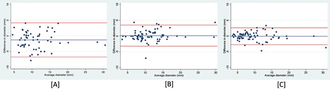
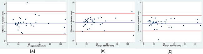
There was a good level of agreement between operator A and operator B in detecting the presence of pelvic endometriotic lesions ( = 0.72). There was a very good level of agreement between operators in identifying endometriotic cysts ( = 0.88) and a good level of agreement in identifying endometriotic nodules ( = 0.61). The inter- and intraobserver repeatability of measuring endometriotic cysts was excellent (intra-class correlation (ICC) ≥ 0.98). There was good interobserver measurement repeatability for bowel nodules (ICC 0.88), but the results for nodules in the posterior compartment were poor (ICC 0.41). The intraobserver repeatability for nodule size measurements was good for both operators (ICC ≥0.86).
Within this cohort, there was insufficient data to perform a separate analysis for nodule size in the anterior compartment. All examinations were performed within a specialised unit with a high prevalence of deep endometriosis. Our findings may not apply to operators without intensive ultrasound training in the diagnosis of pelvic endometriosis.
These findings are important because ultrasound has been widely accepted as the first-line investigation for the diagnosis of pelvic endometriosis, which often determines the need for future investigations and treatment. The detection and measurement of bowel nodules is essential for anticipation of surgical risk and planning surgical excision.
STUDY FUNDING/COMPETING INTERESTS: The authors have no conflict of interest. No funding was obtained for this work.
盆腔超声检测子宫内膜异位病变时,观察者间及观察者内的可重复性如何?
盆腔超声在检测盆腔子宫内膜异位病变方面具有高度可重复性。
经阴道超声(TVS)已被广泛用作盆腔子宫内膜异位症诊断和评估的一线检查方法。超声评估的子宫内膜异位症严重程度与腹腔镜检查显示出良好的一致性(kappa值为0.79)。文献中已描述了TVS使用“滑动征”评估卵巢活动度和道格拉斯窝闭塞情况的可重复性。然而,文献中尚无数据证明对子宫内膜异位囊肿和结节测量的观察者内重复性。
研究设计、规模、持续时间:这是一项为期12个月的前瞻性观察性横断面研究。我们纳入了50名连续就诊的女性,她们在门诊就诊期间均由两名操作人员(A和B)进行检查。
参与者/材料、设置、方法:该研究在一家专业的子宫内膜异位症中心进行。我们纳入了所有在同一次门诊就诊期间由两名经验丰富的操作人员独立进行超声扫描的连续女性。感兴趣的结果是检测子宫内膜异位病变的观察者间及观察者内可重复性。我们还评估了病变大小测量的重复性。
操作人员A和操作人员B在检测盆腔子宫内膜异位病变的存在方面具有良好的一致性(kappa值 = 0.72)。操作人员在识别子宫内膜异位囊肿方面具有非常好的一致性(kappa值 = 0.88),在识别子宫内膜异位结节方面具有良好的一致性(kappa值 = 0.61)。测量子宫内膜异位囊肿的观察者间及观察者内重复性极佳(组内相关系数(ICC)≥0.98)。肠道结节的观察者间测量重复性良好(ICC 0.88),但后盆腔结节的结果较差(ICC 0.41)。两名操作人员对结节大小测量的观察者内重复性均良好(ICC≥0.86)。
局限性、谨慎的原因:在该队列中,没有足够的数据对前盆腔结节大小进行单独分析。所有检查均在深部子宫内膜异位症患病率较高的专业科室进行。我们的研究结果可能不适用于未接受过盆腔子宫内膜异位症诊断超声强化培训的操作人员。
这些结果很重要,因为超声已被广泛接受为盆腔子宫内膜异位症诊断的一线检查方法,这通常决定了未来检查和治疗的必要性。肠道结节的检测和测量对于预测手术风险和规划手术切除至关重要。
研究资金/利益冲突:作者不存在利益冲突。这项工作未获得资金支持。