Institute for Medical Informatics, Statistics and Epidemiology (IMISE), Leipzig University, Härtelstr. 16-18, D-04107 Leipzig, Germany.
Department of Pathology, Hematopathology Section and Lymph Node Registry, University of Kiel/University Hospital Schleswig-Holstein, Arnold-Heller-Str. 3, Haus 14, D-24105 Kiel, Germany.
Gigascience. 2020 Mar 1;9(3). doi: 10.1093/gigascience/giaa016.
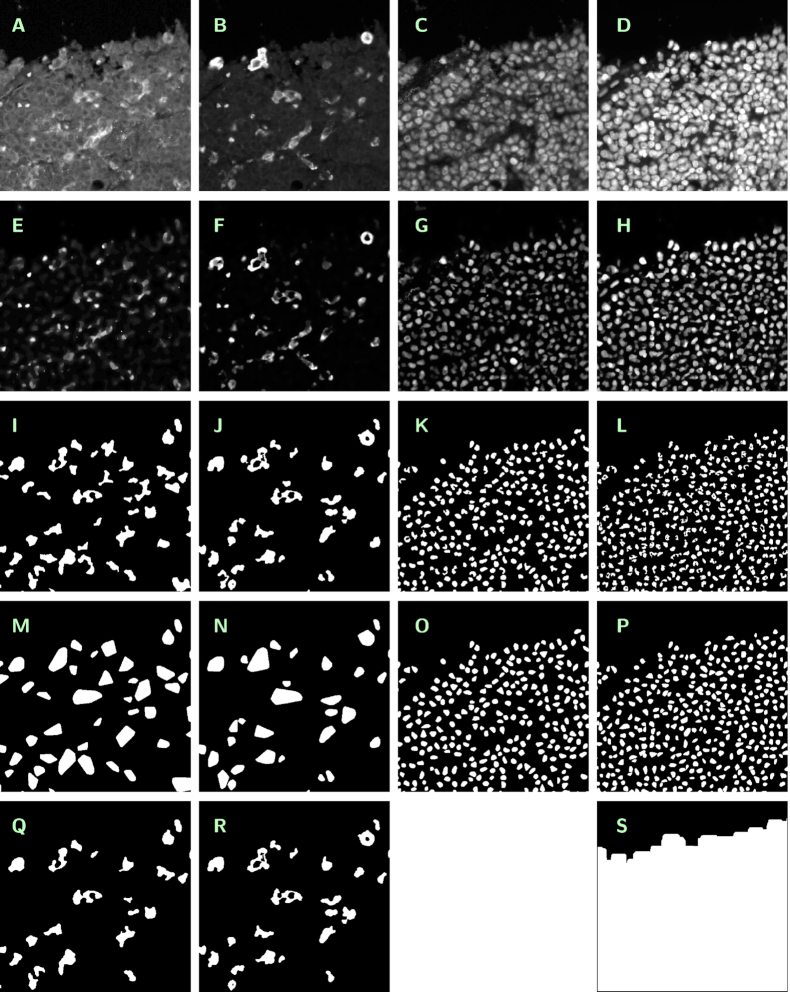
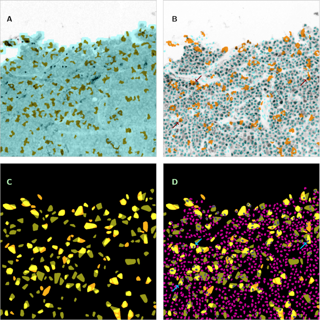
We present an image dataset related to automated segmentation and counting of macrophages in diffuse large B-cell lymphoma (DLBCL) tissue sections. For the classification of DLBCL subtypes, as well as for providing a prognosis of the clinical outcome, the analysis of the tumor microenvironment and, particularly, of the different types and functions of tumor-associated macrophages is indispensable. Until now, however, most information about macrophages has been obtained either in a completely indirect way by gene expression profiling or by manual counts in immunohistochemically (IHC) fluorescence-stained tissue samples while automated recognition of single IHC stained macrophages remains a difficult task. In an accompanying publication, a reliable approach to this problem has been established, and a large set of related images has been generated and analyzed.
Provided image data comprise (i) fluorescence microscopy images of 44 multiple immunohistostained DLBCL tumor subregions, captured at 4 channels corresponding to CD14, CD163, Pax5, and DAPI; (ii) "cartoon-like" total variation-filtered versions of these images, generated by Rudin-Osher-Fatemi denoising; (iii) an automatically generated mask of the evaluation subregion, based on information from the DAPI channel; and (iv) automatically generated segmentation masks for macrophages (using information from CD14 and CD163 channels), B-cells (using information from Pax5 channel), and all cell nuclei (using information from DAPI channel).
A large set of IHC stained DLBCL specimens is provided together with segmentation masks for different cell populations generated by a reference method for automated image analysis, thus featuring considerable reuse potential.
我们提供了一个与弥漫性大 B 细胞淋巴瘤(DLBCL)组织切片中巨噬细胞的自动分割和计数相关的图像数据集。为了对 DLBCL 亚型进行分类,以及为临床结果提供预后,分析肿瘤微环境,特别是不同类型和功能的肿瘤相关巨噬细胞是必不可少的。然而,到目前为止,关于巨噬细胞的大多数信息要么是通过基因表达谱分析以完全间接的方式获得,要么是通过免疫组织化学(IHC)荧光染色组织样本的人工计数获得,而单个 IHC 染色巨噬细胞的自动识别仍然是一个难题。在一篇相关的论文中,已经建立了一种可靠的方法来解决这个问题,并生成和分析了大量相关的图像。
提供的图像数据包括:(i)对应于 CD14、CD163、Pax5 和 DAPI 的 4 个通道的 44 个多个免疫组化染色的 DLBCL 肿瘤亚区的荧光显微镜图像;(ii)通过鲁丁-奥瑟-法蒂米去噪生成的这些图像的“卡通样”全变差滤波版本;(iii)基于 DAPI 通道信息生成的评估子区域的自动生成的掩模;(iv)基于 CD14 和 CD163 通道信息生成的巨噬细胞(使用 CD14 和 CD163 通道信息)、B 细胞(使用 Pax5 通道信息)和所有细胞核(使用 DAPI 通道信息)的自动分割掩模。
提供了大量的 IHC 染色的 DLBCL 标本,并附有通过自动化图像分析的参考方法生成的不同细胞群体的分割掩模,因此具有很大的重用潜力。