Department of Geriatric Medicine (Vascular Medicine), Tokyo Metropolitan Institute of Gerontology, Sakaecho 35-2, Itabashi-ku, Tokyo, 173-0015, Japan.
Cell Commun Signal. 2020 Mar 12;18(1):43. doi: 10.1186/s12964-020-00533-w.
Rapamycin is known to be effective in suppressing senescence and the senescence-associated secretory phenotype (SASP). Therefore, it is highly expected to represent an anti-aging drug. Its anti-aging effect has been demonstrated at the mouse individual level. However, there are not many clinical findings with respect to its activity in humans. Here, we aimed to clarify the effect of rapamycin on human endothelial cells (ECs) as an in vitro model of human blood vessels.
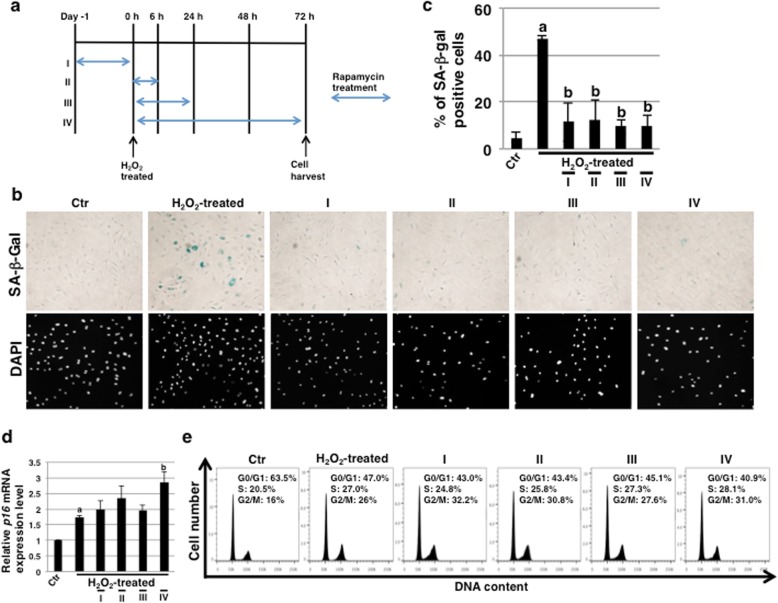
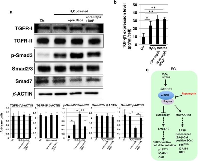
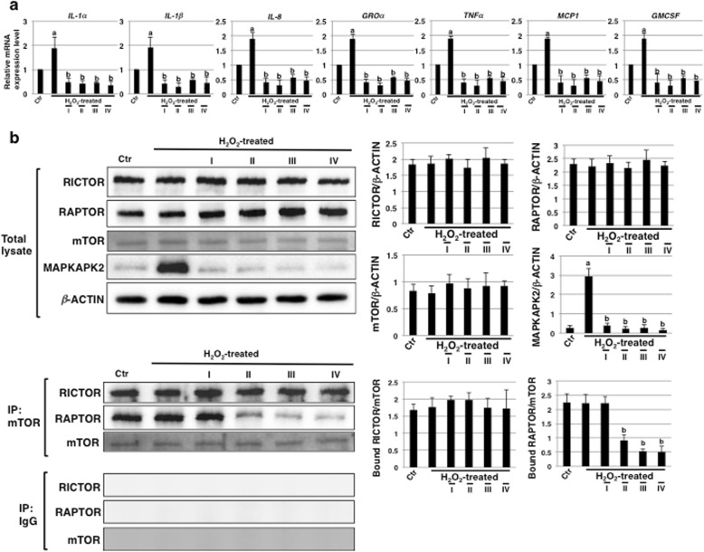
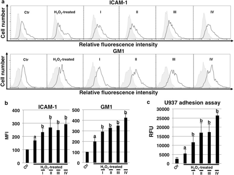
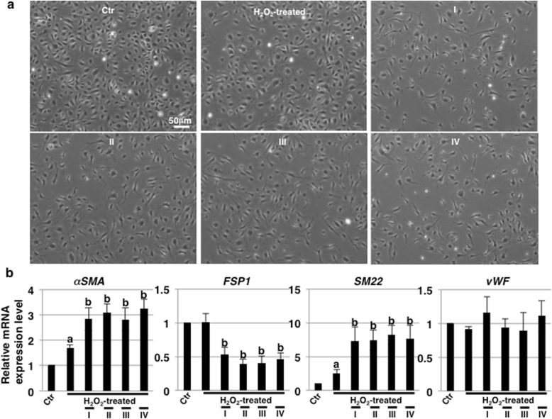
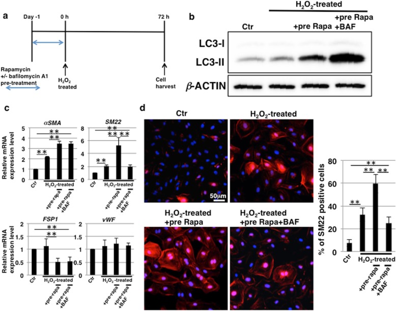
Over the course of oxidative stress-induced senescence using hydrogen peroxide, we examined the effect of rapamycin on human coronary artery ECs (HCAECs). Senescence was evaluated by detecting senescence-associated β-galactosidase (SA-β-Gal) activity and the real-time PCR analysis of p16. Furthermore, expression levels of SASP factors were examined by real-time PCR and the expression of senescence-related antigens, such as intercellular adhesion molecule-1 (ICAM-1) and ganglioside GM1, were examined by fluorescence-activated cell sorting analysis and immunostaining. The inhibitory effect of rapamycin on mTOR signaling was examined by immunoblotting. The adhesion of leukocytes to HCAECs was evaluated by adhesion assays. Endothelial-mesenchymal transition (EndMT) induced by rapamycin treatment was evaluated by real-time PCR analysis and immunostaining for EndMT markers. Finally, we checked the activation of autophagy by immunoblotting and examined its contribution to EndMT by using a specific inhibitor. Furthermore, we examined how the activation of autophagy influences TGF-β signaling by immunoblotting for Smad2/3 and Smad7.
A decrease in SA-β-Gal activity and the suppression of SASP factors were observed in HCAECs undergoing stress-induced premature senescence (SIPS) after rapamycin treatment. In contrast, ICAM-1 and ganglioside GM1 were upregulated by rapamycin treatment. In addition, leukocyte adhesion to HCAECs was promoted by this treatment. In rapamycin-treated HCAECs, morphological changes and the promotion of EndMT were also observed. Furthermore, we found that autophagy activation induced by rapamycin treatment, which led to activation of the TGF-β pathway, contributed to EndMT induction.
We revealed that although rapamycin functions to inhibit senescence and suppress SASP in HCAECs undergoing SIPS, EndMT is induced due to the activation of autophagy. Video abstract.
雷帕霉素在抑制衰老和衰老相关分泌表型(SASP)方面已被证实有效。因此,它有望成为一种抗衰老药物。其抗衰老作用已在小鼠个体水平上得到证实。然而,关于其在人类中的活性的临床发现并不多。在这里,我们旨在阐明雷帕霉素对人内皮细胞(ECs)的作用,作为人类血管的体外模型。
通过使用过氧化氢诱导氧化应激来加速衰老,我们检测了雷帕霉素对人冠状动脉内皮细胞(HCAECs)的影响。通过检测衰老相关β-半乳糖苷酶(SA-β-Gal)活性和实时 PCR 分析 p16 来评估衰老。此外,通过实时 PCR 检测 SASP 因子的表达水平,并通过荧光激活细胞分选分析和免疫染色检测衰老相关抗原,如细胞间黏附分子-1(ICAM-1)和神经节苷脂 GM1 的表达。通过免疫印迹检测雷帕霉素对 mTOR 信号的抑制作用。通过黏附实验评估白细胞与 HCAECs 的黏附。通过实时 PCR 分析和 EndMT 标志物的免疫染色评估雷帕霉素处理诱导的内皮-间充质转化(EndMT)。最后,我们通过免疫印迹检查自噬的激活,并使用特异性抑制剂检查其对 EndMT 的贡献。此外,我们通过免疫印迹检查自噬的激活如何影响 TGF-β 信号,并通过 Smad2/3 和 Smad7 的免疫印迹检查其对 Smad7 的影响。
雷帕霉素处理后,经历应激诱导的过早衰老(SIPS)的 HCAECs 中 SA-β-Gal 活性降低,SASP 因子受到抑制。相反,雷帕霉素处理后 ICAM-1 和神经节苷脂 GM1 上调。此外,白细胞与 HCAECs 的黏附也被这种处理所促进。在雷帕霉素处理的 HCAECs 中,还观察到形态变化和 EndMT 的促进。此外,我们发现雷帕霉素处理诱导的自噬激活导致 TGF-β 途径的激活,有助于 EndMT 的诱导。
我们揭示了尽管雷帕霉素在 SIPS 期间抑制 HCAECs 的衰老并抑制 SASP,但自噬的激活会导致 EndMT 的诱导。