Ankara University Faculty of Medicine, Department of Ophthalmology, Ankara, Turkey
Turk J Ophthalmol. 2020 Mar 5;50(1):31-36. doi: 10.4274/tjo.galenos.2019.20633.
To report the clinical and demographic characteristics, imaging findings, treatment results, and follow-up data of patients with iris cysts.
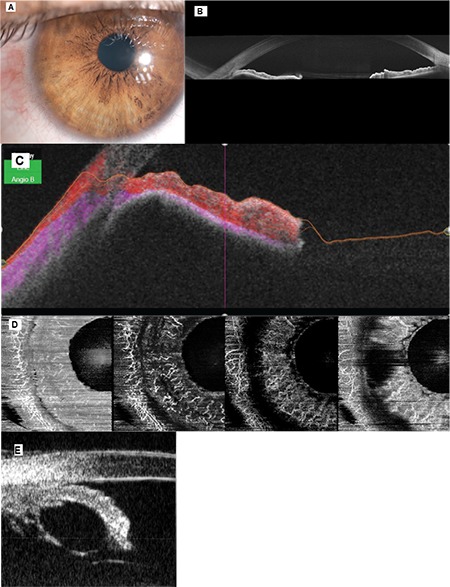
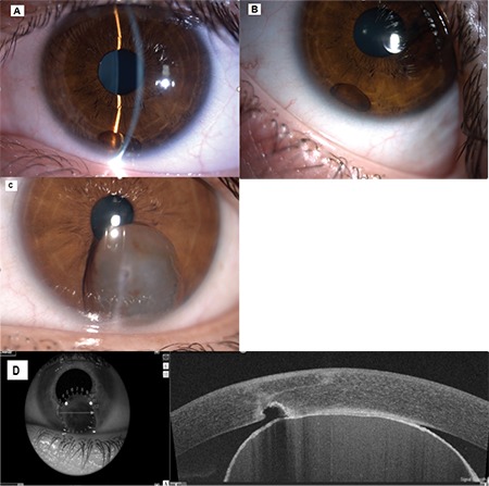
The medical records of 37 patients with iris cysts were retrospectively analyzed. Ultrasound biomicroscopy (UBM), swept-source optical coherence tomography (SS-OCT), and SS-OCT angiography (SS-OCTA) were performed to examine the iris cysts.
The mean age of the patients was 34.4 years, ranging from 5 to 85 years. Twenty-four patients (65%) were female and 13 (35%) were male. Mean follow-up period was 21.3 months, ranging from 4 months to 8 years. Thirty-five (94.5%) of the cysts were classified as primary and 2 (4.5%) were classified as secondary. Thirty-one (83.7%) of the primary cysts were pigment epithelial and 4 were stromal. Primary iris pigment epithelial (IPE) cysts were classified as peripheral in 26 patients (72.2%), midzonal in 4 (11.1%), and dislodged in 1 (2.7%). Stromal cysts were classified as acquired in 3 patients (8.1%) and congenital in 1 patient (2.7%). Secondary iris cysts were caused by perforating eye injury. UBM could visualize both the anterior and posterior surfaces of the cysts (26 patients). Anterior segment SS-OCT could visualize the anterior but not the posterior surface of the cysts (4 patients). Iris cysts did not display intrinsic vascularity on SS-OCTA (4 patients). All pigment epithelial cysts were managed by observation. Of the 4 primary stromal cysts, 3 were managed by surgical excision and 1 by observation. Two secondary cysts required surgical removal.
Pigment epithelial cysts generally remain stable without need for treatment. However, iris stromal cysts frequently require surgical intervention. UBM and SS-OCT were valuable in the diagnosis of iris cysts. On UBM, iris cysts appear with a thin, hyperechoic wall with hypoechoic internal content. Iris cysts did not have intrinsic vascularity on anterior segment SS-OCTA.
报告虹膜囊肿患者的临床和人口统计学特征、影像学表现、治疗结果和随访数据。
回顾性分析了 37 例虹膜囊肿患者的病历。使用超声生物显微镜(UBM)、扫频源光学相干断层扫描(SS-OCT)和 SS-OCT 血管造影(SS-OCTA)检查虹膜囊肿。
患者的平均年龄为 34.4 岁,年龄范围为 5 至 85 岁。24 例(65%)为女性,13 例(35%)为男性。平均随访时间为 21.3 个月,随访时间为 4 个月至 8 年。35 例(94.5%)囊肿为原发性,2 例(4.5%)为继发性。31 例(83.7%)原发性囊肿为色素上皮型,4 例为基质型。原发性虹膜色素上皮(IPE)囊肿在 26 例患者中(72.2%)为周边型,在 4 例患者中(11.1%)为中周型,在 1 例患者中(2.7%)为移位型。基质型囊肿在 3 例患者中(8.1%)为获得性,在 1 例患者中(2.7%)为先天性。继发性虹膜囊肿是由穿透性眼外伤引起的。UBM 可以同时显示囊肿的前后面(26 例)。前节 SS-OCT 可以显示囊肿的前表面,但不能显示后表面(4 例)。SS-OCTA 显示虹膜囊肿无固有血管性(4 例)。所有色素上皮囊肿均通过观察进行治疗。4 例原发性基质囊肿中,3 例通过手术切除,1 例通过观察进行治疗。2 例继发性囊肿需要手术切除。
色素上皮囊肿通常保持稳定,无需治疗。然而,虹膜基质囊肿常需要手术干预。UBM 和 SS-OCT 对虹膜囊肿的诊断具有重要价值。在 UBM 上,虹膜囊肿显示为薄的、高回声壁,伴低回声内部内容物。虹膜囊肿在前节 SS-OCTA 上无固有血管性。