Sarra Angelo, Celluzzi Antonella, Bruno Stefania Paola, Ricci Caterina, Sennato Simona, Ortore Maria Grazia, Casciardi Stefano, Del Chierico Federica, Postorino Paolo, Bordi Federico, Masotti Andrea
Department of Science, University of Roma Tre, Rome, Italy.
Research Laboratories, Bambino Gesù Children's Hospital, IRCCS, Rome, Italy.
Front Microbiol. 2020 Feb 27;11:290. doi: 10.3389/fmicb.2020.00290. eCollection 2020.
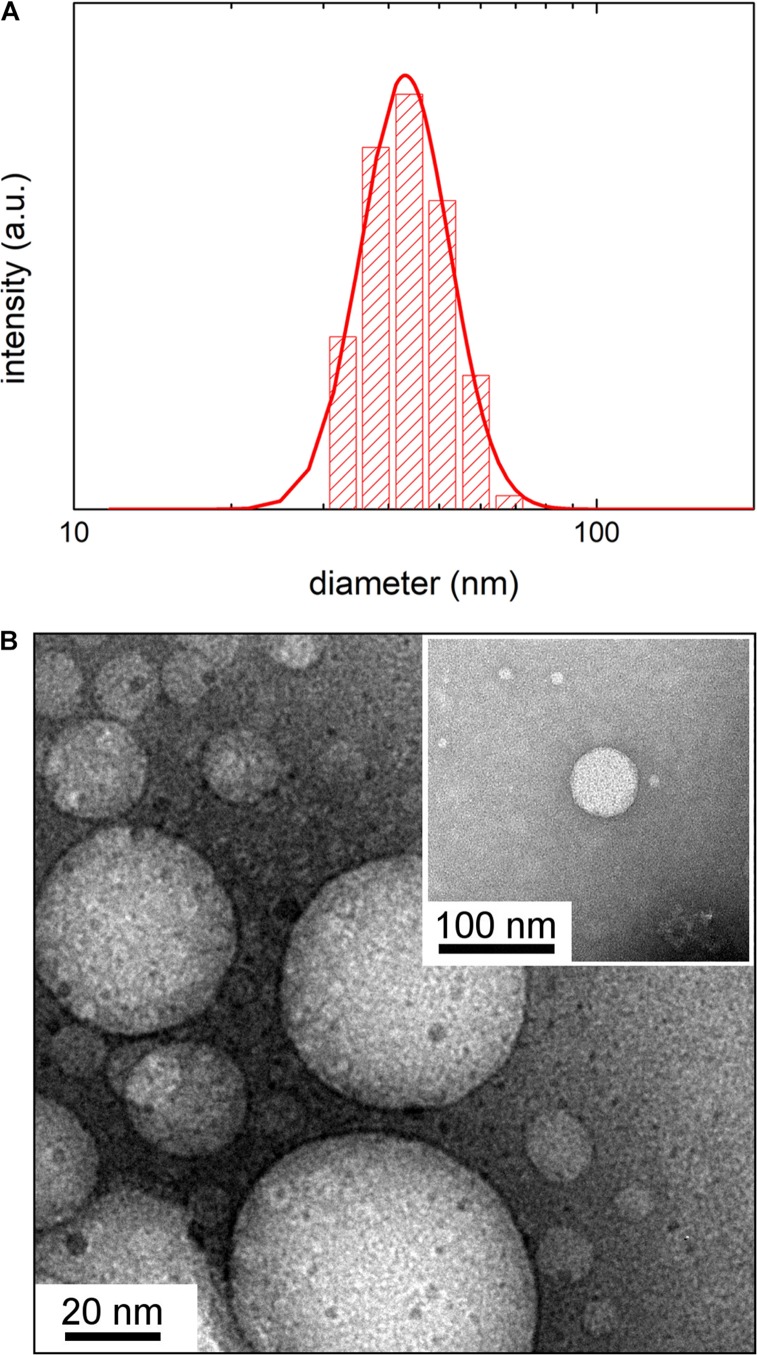
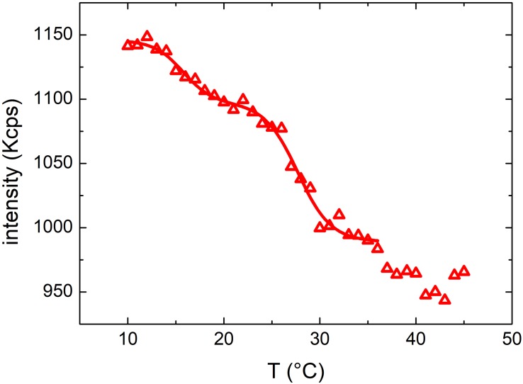
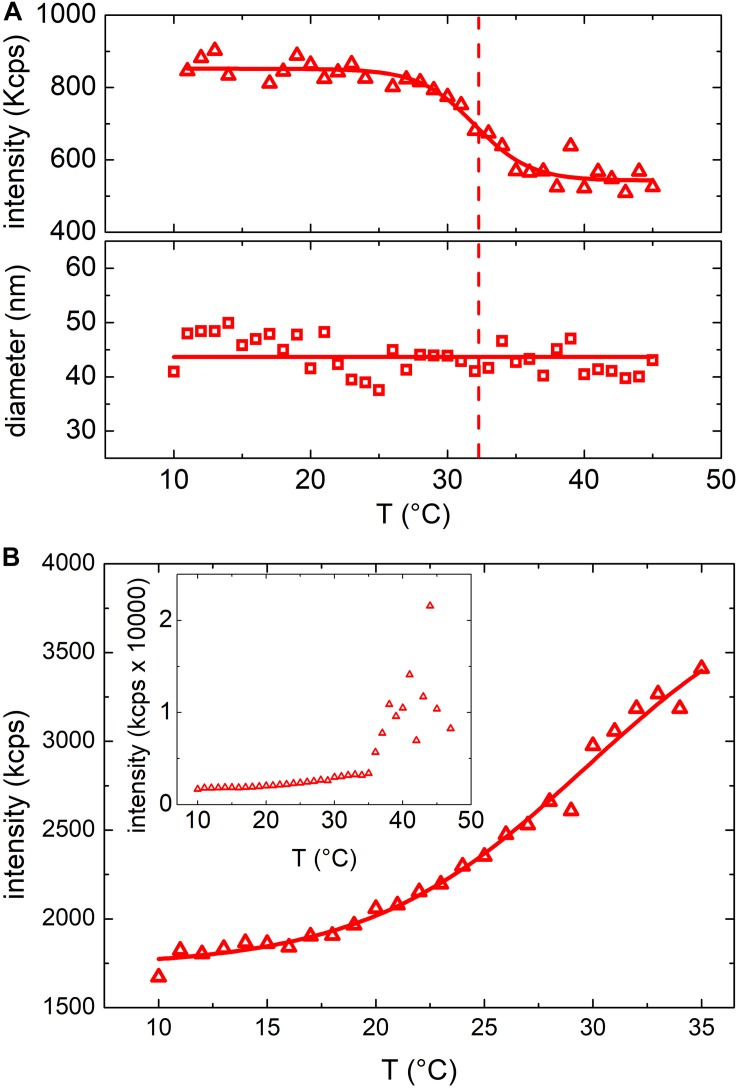
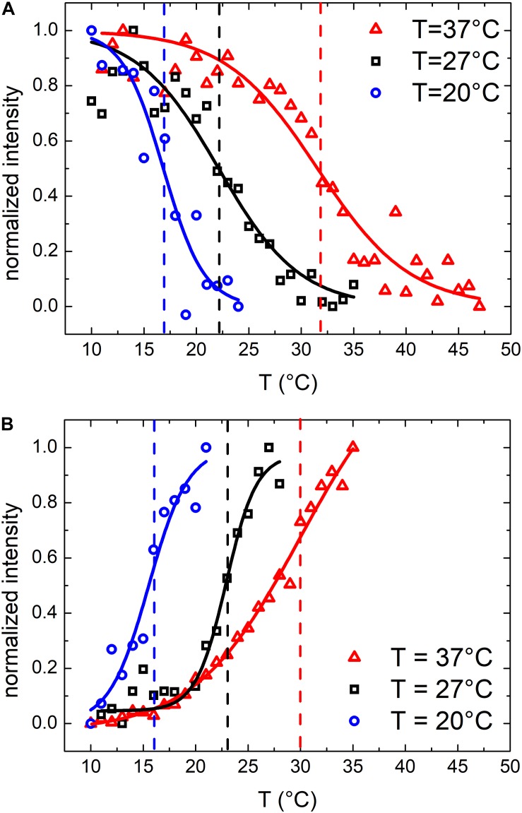
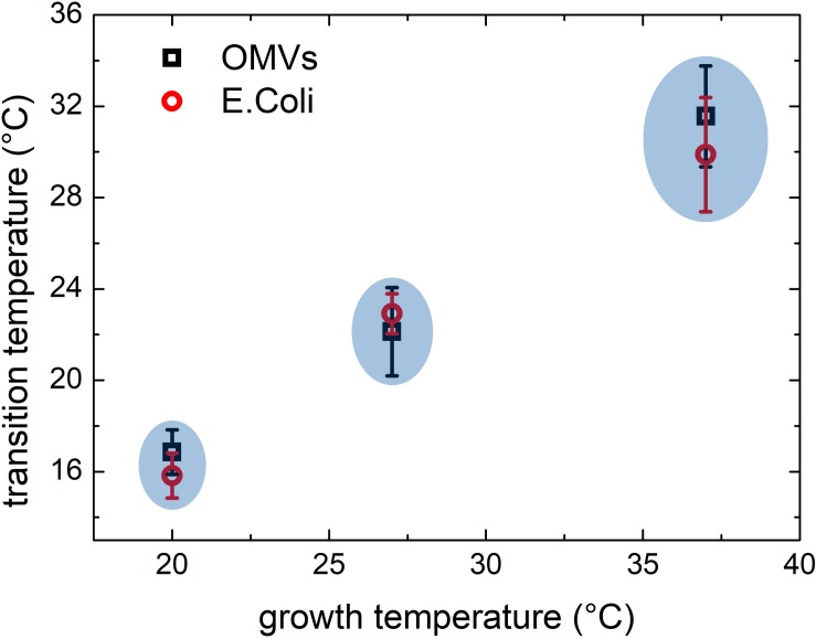
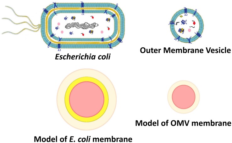
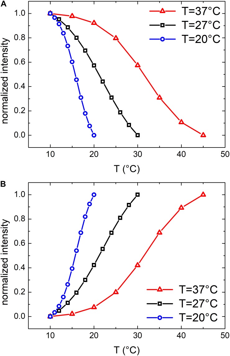
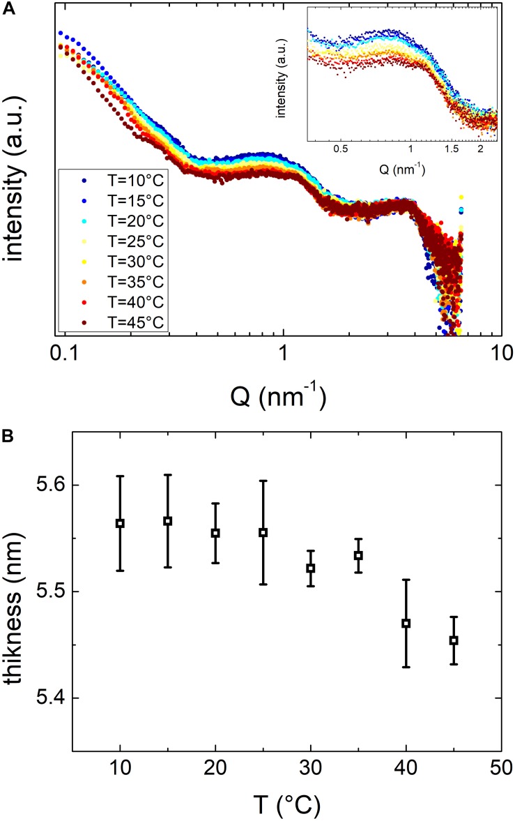
Dynamic Light Scattering (DLS), Small Angle X-ray Scattering (SAXS) and Transmission Electron Microscopy (TEM) are physical techniques widely employed to characterize the morphology and the structure of vesicles such as liposomes or human extracellular vesicles (exosomes). Bacterial extracellular vesicles are similar in size to human exosomes, although their function and membrane properties have not been elucidated in such detail as in the case of exosomes. Here, we applied the above cited techniques, in synergy with the thermotropic characterization of the vesicles lipid membrane using a turbidimetric technique to the study of vesicles produced by Gram-negative bacteria (Outer Membrane Vesicles, OMVs) grown at different temperatures. This study demonstrated that our combined approach is useful to discriminate vesicles of different origin or coming from bacteria cultured under different experimental conditions. We envisage that in a near future the techniques employed in our work will be further implemented to discriminate complex mixtures of bacterial vesicles, thus showing great promises for biomedical or diagnostic applications.
动态光散射(DLS)、小角X射线散射(SAXS)和透射电子显微镜(TEM)是广泛用于表征脂质体或人类细胞外囊泡(外泌体)等囊泡的形态和结构的物理技术。细菌细胞外囊泡的大小与人类外泌体相似,尽管它们的功能和膜特性尚未像外泌体那样得到如此详细的阐明。在这里,我们将上述技术与使用比浊技术对囊泡脂质膜进行热致表征相结合,用于研究在不同温度下生长的革兰氏阴性细菌产生的囊泡(外膜囊泡,OMVs)。这项研究表明,我们的联合方法有助于区分不同来源或来自在不同实验条件下培养的细菌的囊泡。我们设想,在不久的将来,我们工作中使用的技术将进一步完善,以区分细菌囊泡的复杂混合物,从而在生物医学或诊断应用中显示出巨大的前景。