Grande Rossella, Celia Christian, Mincione Gabriella, Stringaro Annarita, Di Marzio Luisa, Colone Marisa, Di Marcantonio Maria C, Savino Luca, Puca Valentina, Santoliquido Roberto, Locatelli Marcello, Muraro Raffaella, Hall-Stoodley Luanne, Stoodley Paul
Department of Pharmacy, "G. d'Annunzio" University of Chieti-PescaraChieti, Italy.
Center of Aging Sciences and Translational MedicineChieti, Italy.
Front Microbiol. 2017 Jun 13;8:1040. doi: 10.3389/fmicb.2017.01040. eCollection 2017.
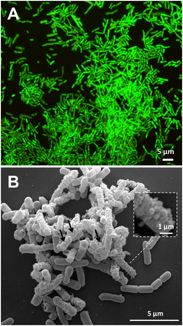
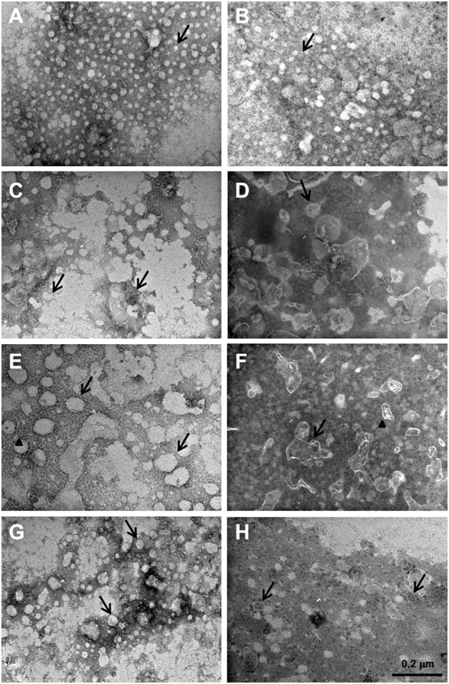
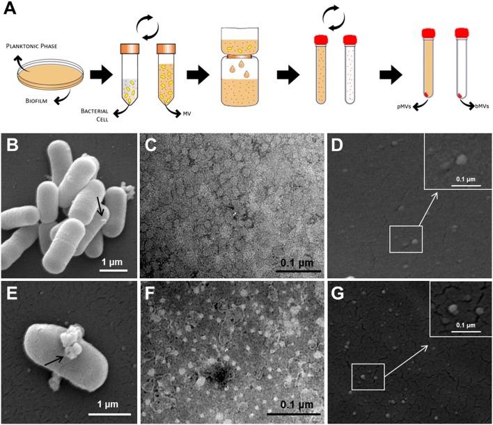
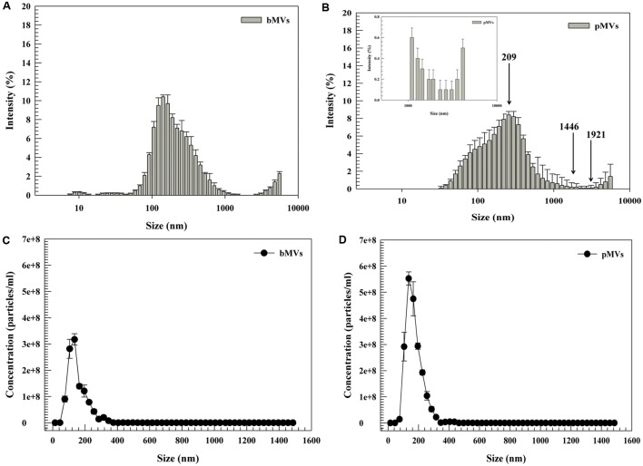
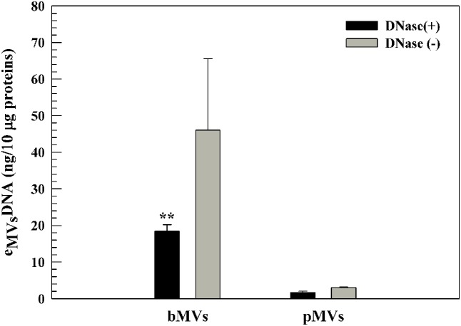
Membrane vesicles (MVs) are bilayer structures which bleb from bacteria, and are important in trafficking biomolecules to other bacteria or host cells. There are few data about MVs produced by the Gram-positive commensal-derived probiotic ; however, MVs from this species may have potential therapeutic benefit. The aim of this study was to detect and characterize MVs produced from biofilm (bMVs), and planktonic (pMVs) phenotypes of DSM 17938. MVs were analyzed for structure and physicochemical characterization by Scanning Electron Microscope (SEM) and Dynamic Light Scattering (DLS). Their composition was interrogated using various digestive enzyme treatments and subsequent Transmission Electron Microscopy (TEM) analysis. eDNA (extracellular DNA) was detected and quantified using PicoGreen. We found that planktonic and biofilm of cultures generated MVs with a broad size distribution. Our data also showed that eDNA was associated with pMVs and bMVs (eDNA). DNase I treatment demonstrated no modifications of MVs, suggesting that an eDNA-MVs complex protected the eDNA. Proteinase K and Phospholipase C treatments modified the structure of MVs, showing that lipids and proteins are important structural components of MVs. The biological composition and the physicochemical characterization of MVs generated by the probiotic may represent a starting point for future applications in the development of vesicles-based therapeutic systems.
膜泡(MVs)是从细菌表面膨出的双层结构,在将生物分子运输到其他细菌或宿主细胞中起着重要作用。关于革兰氏阳性共生来源益生菌产生的膜泡的数据很少;然而,该物种产生的膜泡可能具有潜在的治疗益处。本研究的目的是检测和表征DSM 17938生物膜(bMVs)和浮游(pMVs)表型产生的膜泡。通过扫描电子显微镜(SEM)和动态光散射(DLS)对膜泡进行结构和物理化学表征分析。使用各种消化酶处理并随后进行透射电子显微镜(TEM)分析来探究其组成。使用PicoGreen检测和定量细胞外DNA(eDNA)。我们发现培养物的浮游和生物膜产生了大小分布广泛的膜泡。我们的数据还表明,eDNA与pMVs和bMVs(eDNA)相关。DNA酶I处理未显示膜泡有任何改变,表明eDNA-膜泡复合物保护了eDNA。蛋白酶K和磷脂酶C处理改变了膜泡的结构,表明脂质和蛋白质是膜泡的重要结构成分。益生菌产生的膜泡的生物学组成和物理化学表征可能代表了基于膜泡的治疗系统未来开发应用的起点。