College of Life Science, Qingdao Agricultural University, No. 700, Changcheng Road, Chengyang District, Qingdao, 266109, Shandong, People's Republic of China.
Department of Surgery, Medical University of South Carolina, Charleston, SC, 29425, USA.
Stem Cell Res Ther. 2020 Mar 17;11(1):120. doi: 10.1186/s13287-020-01621-x.
Adipose-derived mesenchymal stem cells (ASCs) therapy is emerging as a novel therapeutic option for the treatment of a variety of diseases including diabetes and diabetic wound healing. Multiple studies indicate that ASCs could promote wound healing and reverse diabetes. However, whether ASCs from diabetic donors retain their therapeutic functions and the mechanisms of how ASCs contribute to wound healing remain largely unknown. In this study, we explored the cutaneous wound healing ability of ASCs collected from C57BL/6 mice that had been rendered diabetic with streptozotocin (STZ).
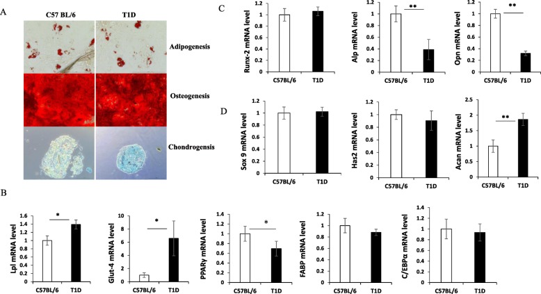
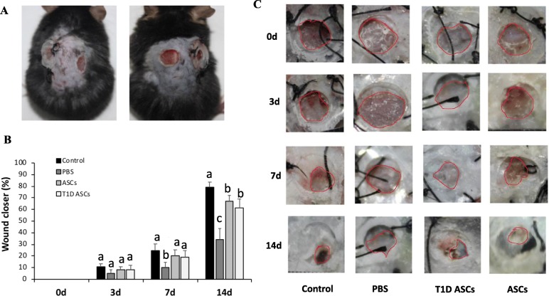
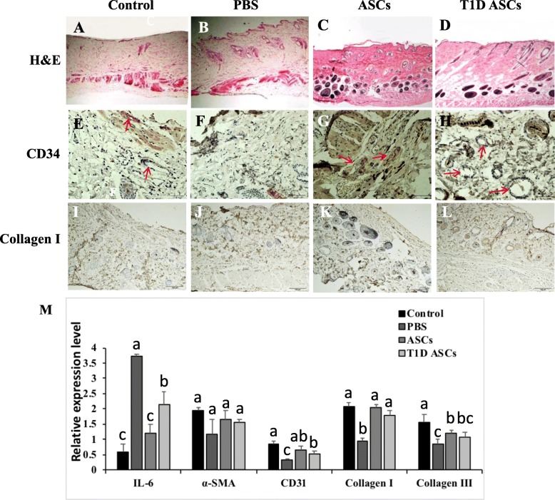
ASCs were harvested from adipose tissues of type 1 diabetic (T1D) or normal C57BL/6 mice. Cell phenotypes were evaluated by flow cytometry analysis, and cell differentiation into adipocytes, chondrocytes, and osteocytes was compared. Secretions of transforming growth factor β (TGF-β1), basic fibroblast growth factor (bFGF), and vascular endothelial growth factor (VEGF) by ASCs were assessed by ELISA. Migration and proliferation of fibroblasts co-cultured with T1D ASCs or control ASCs were also compared. The therapeutic effects of T1D and control ASCs in promoting wound closure were measured in vivo in a T1D wound mouse model. Granulation tissues were collected and stained with H&E at 14th day. CD34 and collagen I were detected by immunohistochemistry. Expressions of IL-6, α-SMA, CD31, collagen I, and collagen III were quantified by real-time PCR. GFP-expressing ASCs were used to trace in vivo cell differentiation.
T1D ASCs and control ASCs showed similar expression of cell surface markers (CD29, CD34, CD105) and proliferation pattern. They can both differentiate into different cell types. T1D ASCs secreted similar amounts of VEGF and bFGF, but less TGF-β compared with control ASCs. Like control ASCs, T1D ASCs promoted the proliferation and migration of skin fibroblast cells. When injected in cutaneous wound of T1D mice, T1D ASCs increased wound closure and hair follicle regeneration at a comparable extent as ASCs. Mice receiving T1D ASCs or ASCs exhibited significantly higher expressions of collagen I, collagen III, and CD31 and reduced expression of IL-6 in wound tissues. Immunohistochemistry staining showed increased angiogenesis in mice receiving ASCs as was evident by increased CD34 cells and collagen I staining. GFP ASCs injection showed that ASCs differentiated into fibroblasts and endothelial cells in vivo.
Our results suggest that T1D ASCs could accelerate cutaneous wound healing. Mechanisms may include increasing fibroblast growth and migration, skin angiogenesis, and differentiation into fibroblasts and endothelial cells. This study provides evidence that diabetic ASCs may be used as a therapeutic option in cutaneous wound healing in diabetic recipients.
脂肪间充质干细胞(ASCs)治疗作为一种治疗多种疾病的新方法正在出现,包括糖尿病和糖尿病创面愈合。多项研究表明,ASCs 可以促进创面愈合并逆转糖尿病。然而,糖尿病供体来源的 ASCs 是否保留其治疗功能,以及 ASCs 如何促进创面愈合的机制在很大程度上仍不清楚。在这项研究中,我们探讨了从小鼠脂肪组织中提取的 ASC 在链脲佐菌素(STZ)致糖尿病的 C57BL/6 小鼠中的皮肤创面愈合能力。
从 1 型糖尿病(T1D)或正常 C57BL/6 小鼠的脂肪组织中提取 ASC。通过流式细胞术分析评估细胞表型,并比较细胞向脂肪细胞、软骨细胞和成骨细胞的分化。通过 ELISA 评估 ASC 分泌转化生长因子β(TGF-β1)、碱性成纤维细胞生长因子(bFGF)和血管内皮生长因子(VEGF)的情况。还比较了与 T1D ASC 或对照 ASC 共培养的成纤维细胞的迁移和增殖。通过在 T1D 创面小鼠模型中测量体内 T1D 和对照 ASC 促进创面闭合的效果来评估其治疗效果。在第 14 天收集肉芽组织并进行 H&E 染色。通过免疫组织化学检测 CD34 和胶原 I。通过实时 PCR 定量检测白细胞介素 6(IL-6)、α-SMA、CD31、胶原 I 和胶原 III 的表达。使用 GFP 表达的 ASC 来追踪体内细胞分化。
T1D ASC 和对照 ASC 均表现出相似的细胞表面标志物(CD29、CD34、CD105)表达和增殖模式。它们都可以分化为不同的细胞类型。T1D ASC 分泌的 VEGF 和 bFGF 量相似,但 TGF-β 分泌量低于对照 ASC。与对照 ASC 一样,T1D ASC 促进皮肤成纤维细胞的增殖和迁移。当注射到 T1D 小鼠的皮肤创面上时,T1D ASC 与 ASC 一样,可增加创面闭合和毛囊再生的程度。接受 T1D ASC 或 ASC 的小鼠在创面组织中表现出更高的胶原 I、胶原 III 和 CD31 表达,以及更低的白细胞介素 6 表达。免疫组织化学染色显示,接受 ASC 的小鼠血管生成增加,这表现在 CD34 细胞和胶原 I 染色增加。GFP ASC 注射显示 ASC 在体内分化为成纤维细胞和内皮细胞。
我们的研究结果表明,T1D ASC 可以加速皮肤创面愈合。其机制可能包括增加成纤维细胞的生长和迁移、皮肤血管生成以及分化为成纤维细胞和内皮细胞。这项研究提供了证据表明,糖尿病来源的 ASC 可作为糖尿病受者皮肤创面愈合的治疗选择。