Wankhade Archana B, Patro Priyadarshini, Arora Ripu Daman, Nagarkar Nitin M
Department of Microbiology, AIIMS Raipur, Raipur, Chhattisgarh, India.
Department of ENT, Head and Neck Surgery, AIIMS Raipur, Raipur, Chhattisgarh, India.
J Oral Maxillofac Pathol. 2020 Feb;24(Suppl 1):S124-S127. doi: 10.4103/jomfp.JOMFP_356_19. Epub 2020 Feb 28.
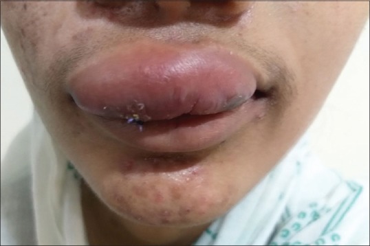
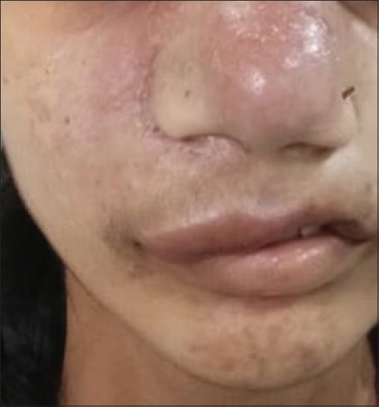
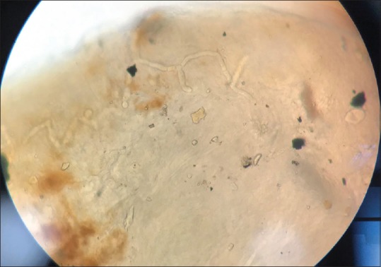
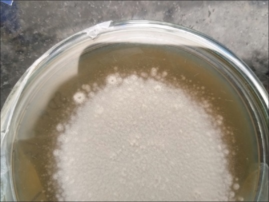
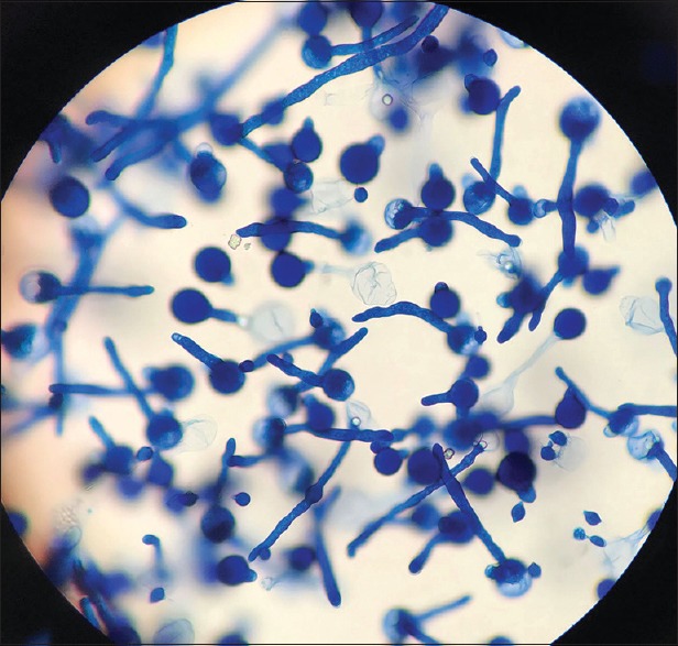
Rhinoentomophthoromycosis due to is a rare, chronic, granulomatous disease, occurring mainly in tropical countries including India. We report two cases of rhinoentomophthoromycosis in an 18-year-old female and a farmer of 35 years residents of Chhattisgarh shifted from Madhya Pradesh and Orrisa. It was diagnosed by microscopy and isolation in culture. The patient presented with a swollen nose with obstruction that had progressed slowly over 1 year. His nasal swelling was bilateral, diffuse, mildly tender, erythematous, nonpitting, with mucosal crusting and hypertrophy of inferior turbinates but no regional lymphadenopathy. Culture of tissue from the nasal biopsy on sabouraud dextrose agar yielded multiple colonies of a mold with satellite smaller colonies at periphery. The isolate demonstrated the macroscopic and microscopic morphologic characteristics of . The patients were earlier treated with itraconazole or its combination with potassium iodide and the patients were treated successfully with amphotericin B.
由[病原体名称未给出]引起的鼻虫霉病是一种罕见的慢性肉芽肿性疾病,主要发生在包括印度在内的热带国家。我们报告了两例鼻虫霉病病例,一例是一名18岁女性,另一例是一名35岁的农民,他们都是从中央邦和奥里萨邦迁移至恰蒂斯加尔邦的居民。该病通过显微镜检查和培养分离进行诊断。患者表现为鼻子肿胀并伴有阻塞,这种情况在1年多的时间里缓慢进展。其鼻肿胀为双侧、弥漫性,轻度压痛,呈红斑样,非凹陷性,伴有黏膜结痂和下鼻甲肥大,但无局部淋巴结肿大。在沙氏葡萄糖琼脂上对鼻活检组织进行培养,产生了多个霉菌菌落,周边有卫星状较小菌落。分离株显示出[病原体名称未给出]的宏观和微观形态特征。患者早期接受伊曲康唑治疗或其与碘化钾联合治疗,之后使用两性霉素B成功治愈。