Department of Gastroenterology, Affiliated Hospital of Jining Medical University, Jining, Shandong, China (mainland).
Department of Spine Surgery, Affiliated Hospital of Jining Medical University, Jining, Shandong, China (mainland).
Med Sci Monit. 2020 Mar 19;26:e921887. doi: 10.12659/MSM.921887.
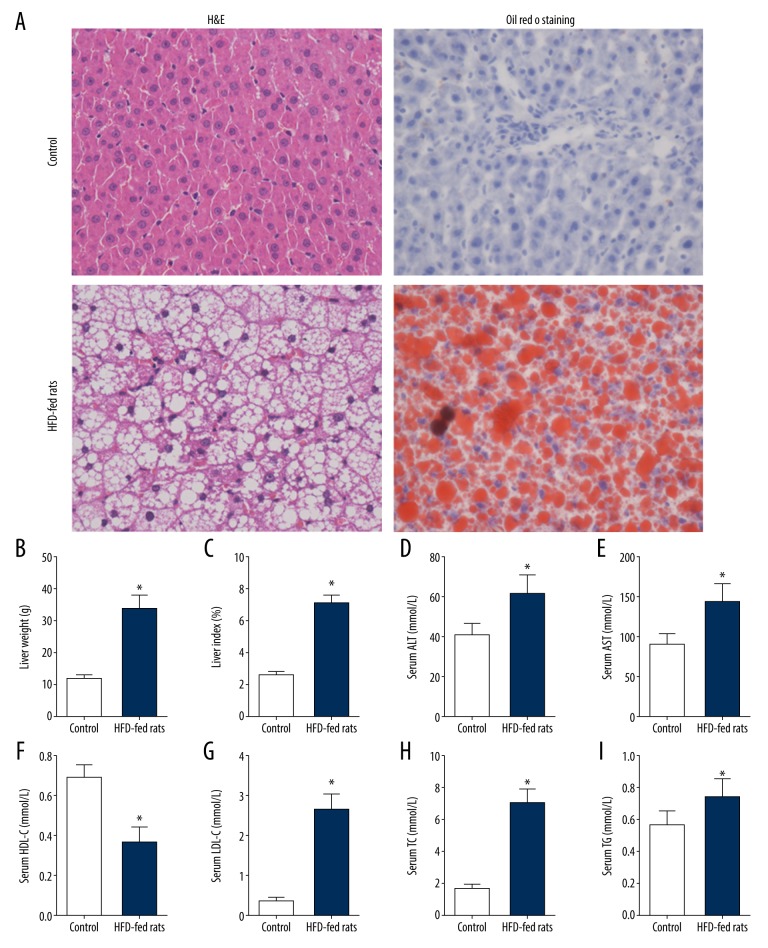
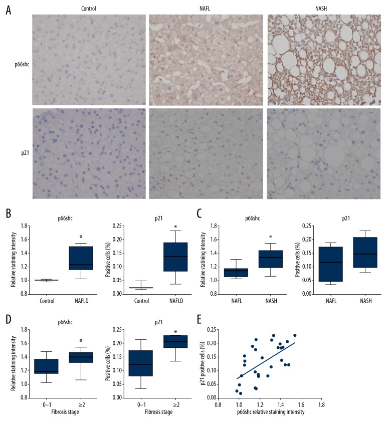
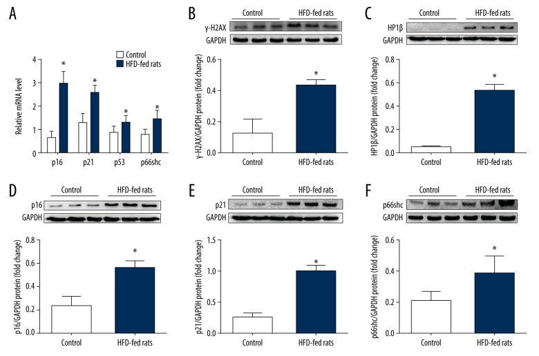
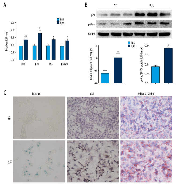
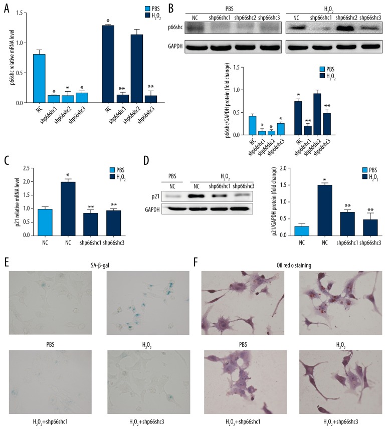
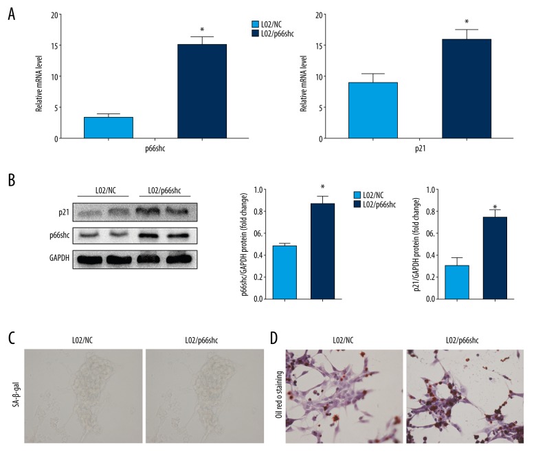
BACKGROUND Recent studies have suggested that hepatocyte senescence could contribute to hepatic steatosis and its progression in nonalcoholic fatty liver disease (NAFLD). However, the underlying mechanism causing hepatocyte senescence in this pathological condition is still unclear. A thorough understanding of the mechanism could provide a new target for therapeutic intervention. The purpose of this study was to investigate the role of p66shc in hepatocyte senescence and hepatocyte damage in NAFLD progression. MATERIAL AND METHODS We examined the expression levels of hepatic p66shc and senescence markers in rats and humans with NAFLD, and we assessed the effect of p66shc knockdown or overexpression on senescence and steatosis in human liver cells. RESULTS In this study, we showed that increased hepatic p66shc expression was consistent with upregulated expression of the following senescence markers in NAFLD rats: heterochromatin protein-1-beta (HP1ß), p16, p21, and p53. Furthermore, senescence and steatosis could be induced in hepatoblastoma cell line (HepG2) cells when cells were stimulated with a low concentration of H₂O₂, and this effect was significantly alleviated by knockdown of p66shc. However, overexpression of p66shc could promote senescence and steatosis in L02 cells. Finally, increased hepatic p66shc protein levels correlated with enhanced expression of the senescence marker p21 and mirrored the degree of disease severity in NAFLD patients. CONCLUSIONS Our findings indicated that the increase in hepatocyte senescence and steatosis in NAFLD may be caused by the upregulation of p66shc expression, implying that strategies for p66shc-mediated regulation of hepatocyte senescence may provide new therapeutic tools for NAFLD.
最近的研究表明,肝细胞衰老可能导致非酒精性脂肪性肝病(NAFLD)中的肝脂肪变性及其进展。然而,在这种病理条件下导致肝细胞衰老的潜在机制仍不清楚。深入了解该机制可为治疗干预提供新的靶点。本研究旨在探讨 p66shc 在 NAFLD 进展过程中肝细胞衰老和肝细胞损伤中的作用。
我们检测了 NAFLD 大鼠和人类肝脏中的 p66shc 和衰老标志物的表达水平,并评估了 p66shc 敲低或过表达对人肝细胞衰老和脂肪变性的影响。
本研究表明,在 NAFLD 大鼠中,肝 p66shc 表达增加与衰老标志物异染色质蛋白 1-β(HP1ß)、p16、p21 和 p53 的上调表达一致。此外,当用低浓度 H₂O₂刺激肝癌细胞系(HepG2)细胞时,可诱导细胞衰老和脂肪变性,而 p66shc 敲低可显著减轻这种作用。然而,p66shc 的过表达可促进 L02 细胞的衰老和脂肪变性。最后,肝 p66shc 蛋白水平的增加与衰老标志物 p21 的表达增强相关,并反映了 NAFLD 患者疾病严重程度。
我们的研究结果表明,NAFLD 中肝细胞衰老和脂肪变性的增加可能是由 p66shc 表达上调引起的,这表明针对 p66shc 介导的肝细胞衰老的调节策略可能为 NAFLD 提供新的治疗工具。