• 文献检索
  • 文档翻译
  • 深度研究
  • 学术资讯
  • Suppr Zotero 插件Zotero 插件
  • 邀请有礼
  • 套餐&价格
  • 历史记录
应用&插件
Suppr Zotero 插件Zotero 插件浏览器插件Mac 客户端Windows 客户端微信小程序
定价
高级版会员购买积分包购买API积分包
服务
文献检索文档翻译深度研究API 文档MCP 服务
关于我们
关于 Suppr公司介绍联系我们用户协议隐私条款
关注我们

Suppr 超能文献

核心技术专利:CN118964589B侵权必究
粤ICP备2023148730 号-1Suppr @ 2026

文献检索

告别复杂PubMed语法,用中文像聊天一样搜索,搜遍4000万医学文献。AI智能推荐,让科研检索更轻松。

立即免费搜索

文件翻译

保留排版,准确专业,支持PDF/Word/PPT等文件格式,支持 12+语言互译。

免费翻译文档

深度研究

AI帮你快速写综述,25分钟生成高质量综述,智能提取关键信息,辅助科研写作。

立即免费体验

新型冠状病毒病 2019(COVID-19)的胸部 CT 表现:影像学综述。

Chest CT manifestations of new coronavirus disease 2019 (COVID-19): a pictorial review.

机构信息

Department of Radiology, West China Hospital, Sichuan University, No. 37 Guoxue Alley, Wuhou District, Chengdu, 610041, China.

出版信息

Eur Radiol. 2020 Aug;30(8):4381-4389. doi: 10.1007/s00330-020-06801-0. Epub 2020 Mar 19.

DOI:10.1007/s00330-020-06801-0
PMID:32193638
原文链接:https://pmc.ncbi.nlm.nih.gov/articles/PMC7088323/
Abstract

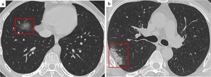
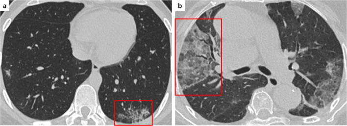
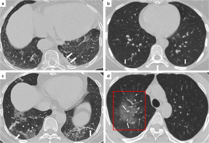
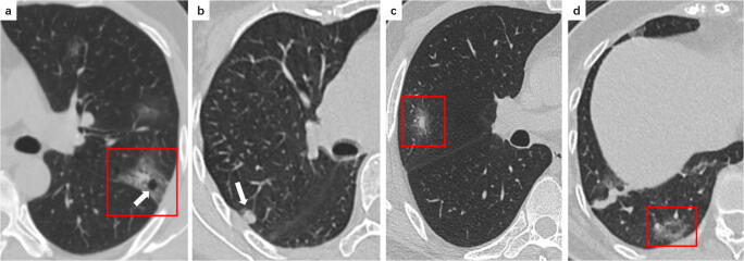
Coronavirus disease 2019 (COVID-19) outbreak, first reported in Wuhan, China, has rapidly swept around the world just within a month, causing global public health emergency. In diagnosis, chest computed tomography (CT) manifestations can supplement parts of limitations of real-time reverse transcription polymerase chain reaction (RT-PCR) assay. Based on a comprehensive literature review and the experience in the frontline, we aim to review the typical and relatively atypical CT manifestations with representative COVID-19 cases at our hospital, and hope to strengthen the recognition of these features with radiologists and help them make a quick and accurate diagnosis.Key Points• Ground glass opacities, consolidation, reticular pattern, and crazy paving pattern are typical CT manifestations of COVID-19.• Emerging atypical CT manifestations, including airway changes, pleural changes, fibrosis, nodules, etc., were demonstrated in COVID-19 patients.• CT manifestations may associate with the progression and prognosis of COVID-19.

摘要

新型冠状病毒病 2019(COVID-19)疫情,首次在中国武汉报告,在短短一个月内迅速席卷全球,引发全球公共卫生紧急事件。在诊断方面,胸部计算机断层扫描(CT)表现可以补充实时逆转录聚合酶链反应(RT-PCR)检测的部分局限性。基于全面的文献复习和一线经验,我们旨在回顾我院具有代表性的 COVID-19 病例的典型和相对非典型 CT 表现,希望增强放射科医生对这些特征的认识,并帮助他们快速准确地诊断。

关键点

  • 磨玻璃影、实变、网状影和铺路石征是 COVID-19 的典型 CT 表现。

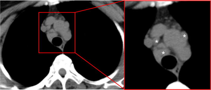
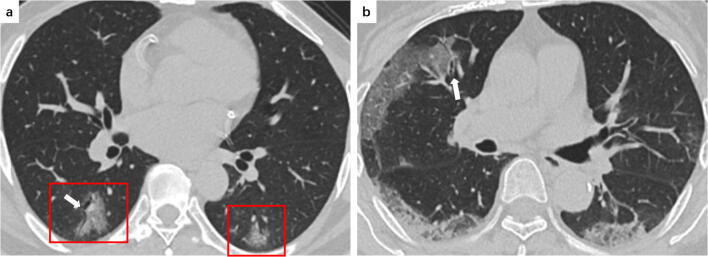
  • COVID-19 患者表现出新兴的非典型 CT 表现,包括气道改变、胸膜改变、纤维化、结节等。

  • CT 表现可能与 COVID-19 的进展和预后相关。

https://cdn.ncbi.nlm.nih.gov/pmc/blobs/356d/7088323/580ea2777345/330_2020_6801_Fig6_HTML.jpg
https://cdn.ncbi.nlm.nih.gov/pmc/blobs/356d/7088323/cbbbec5d209f/330_2020_6801_Fig1_HTML.jpg
https://cdn.ncbi.nlm.nih.gov/pmc/blobs/356d/7088323/3ea3705643c5/330_2020_6801_Fig2_HTML.jpg
https://cdn.ncbi.nlm.nih.gov/pmc/blobs/356d/7088323/9ca4b1e66481/330_2020_6801_Fig3_HTML.jpg
https://cdn.ncbi.nlm.nih.gov/pmc/blobs/356d/7088323/d8cbd39a2c68/330_2020_6801_Fig4_HTML.jpg
https://cdn.ncbi.nlm.nih.gov/pmc/blobs/356d/7088323/137bcb7b299c/330_2020_6801_Fig5_HTML.jpg
https://cdn.ncbi.nlm.nih.gov/pmc/blobs/356d/7088323/580ea2777345/330_2020_6801_Fig6_HTML.jpg

相似文献

1
Chest CT manifestations of new coronavirus disease 2019 (COVID-19): a pictorial review.新型冠状病毒病 2019(COVID-19)的胸部 CT 表现:影像学综述。
Eur Radiol. 2020 Aug;30(8):4381-4389. doi: 10.1007/s00330-020-06801-0. Epub 2020 Mar 19.
2
Imaging and clinical features of patients with 2019 novel coronavirus SARS-CoV-2.新型冠状病毒 2019 年 SARS-CoV-2 患者的影像学和临床特征。
Eur J Nucl Med Mol Imaging. 2020 May;47(5):1275-1280. doi: 10.1007/s00259-020-04735-9. Epub 2020 Feb 28.
3
CT Manifestations and Clinical Characteristics of 1115 Patients with Coronavirus Disease 2019 (COVID-19): A Systematic Review and Meta-analysis.新冠肺炎患者 1115 例的 CT 表现及临床特征:系统评价和荟萃分析。
Acad Radiol. 2020 Jul;27(7):910-921. doi: 10.1016/j.acra.2020.04.033. Epub 2020 May 5.
4
Chest CT features of coronavirus disease 2019 (COVID-19) pneumonia: key points for radiologists.COVID-19 肺炎的胸部 CT 特征:放射科医生的关键点。
Radiol Med. 2020 Jul;125(7):636-646. doi: 10.1007/s11547-020-01237-4. Epub 2020 Jun 4.
5
Performance of Radiologists in Differentiating COVID-19 from Non-COVID-19 Viral Pneumonia at Chest CT.放射科医生在胸部 CT 鉴别 COVID-19 与非 COVID-19 病毒性肺炎中的表现。
Radiology. 2020 Aug;296(2):E46-E54. doi: 10.1148/radiol.2020200823. Epub 2020 Mar 10.
6
An uncommon manifestation of COVID-19 pneumonia on CT scan with small cavities in the lungs: A case report.新冠肺炎肺部CT扫描显示肺部有小空洞的罕见表现:一例报告。
Medicine (Baltimore). 2020 Jul 10;99(28):e21240. doi: 10.1097/MD.0000000000021240.
7
Halo, Reversed Halo, or Both? Atypical Computed Tomography Manifestations of Coronavirus Disease (COVID-19) Pneumonia: The "Double Halo Sign".你好,反晕征还是双晕征?COVID-19 肺炎的非典型计算机断层扫描表现:“双晕征”。
Korean J Radiol. 2020 Oct;21(10):1161-1164. doi: 10.3348/kjr.2020.0687. Epub 2020 Jul 17.
8
Dynamic Chest CT Evaluation in Three Cases of 2019 Novel Coronavirus Pneumonia.动态胸部 CT 评估 2019 年新型冠状病毒肺炎三例
Arch Iran Med. 2020 Apr 1;23(4):277-280. doi: 10.34172/aim.2020.11.
9
Spectrum of chest computed tomographic (CT) findings in coronavirus disease-19 (COVID-19) patients in India.印度 COVID-19 患者的胸部计算机断层扫描(CT)表现谱。
Eur J Radiol. 2020 Aug;129:109147. doi: 10.1016/j.ejrad.2020.109147. Epub 2020 Jun 24.
10
Frequency and Distribution of Chest Radiographic Findings in Patients Positive for COVID-19.COVID-19 阳性患者的胸部 X 线表现的频率和分布。
Radiology. 2020 Aug;296(2):E72-E78. doi: 10.1148/radiol.2020201160. Epub 2020 Mar 27.

引用本文的文献

1
Visual and AI-Based Assessment of COVID-19 Pneumonia: Practicability and Reproducibility of an Established Semi-Quantitative Chest CT Scoring System.基于视觉和人工智能的新型冠状病毒肺炎评估:一种成熟的半定量胸部CT评分系统的实用性和可重复性
Diagnostics (Basel). 2025 Aug 8;15(16):1987. doi: 10.3390/diagnostics15161987.
2
Reversed halo sign: from a clinico-radiologic-pathologic point of view.反晕征:从临床-放射-病理角度分析
Eur J Med Res. 2025 Jun 16;30(1):482. doi: 10.1186/s40001-025-02665-6.
3
Biomarker Patterns and Their Association with Lung Injury in COVID-19 Patients.

本文引用的文献

1
The Many Faces of COVID-19: Spectrum of Imaging Manifestations.新冠病毒肺炎的多样面貌:影像表现谱
Radiol Cardiothorac Imaging. 2020 Feb 14;2(1):e200037. doi: 10.1148/ryct.2020200037. eCollection 2020 Feb.
2
Imaging Profile of the COVID-19 Infection: Radiologic Findings and Literature Review.新型冠状病毒肺炎感染的影像学表现:放射学发现与文献综述
Radiol Cardiothorac Imaging. 2020 Feb 13;2(1):e200034. doi: 10.1148/ryct.2020200034. eCollection 2020 Feb.
3
Severe Acute Respiratory Disease in a Huanan Seafood Market Worker: Images of an Early Casualty.
新型冠状病毒肺炎患者的生物标志物模式及其与肺损伤的关联
Medicina (Kaunas). 2025 May 21;61(5):931. doi: 10.3390/medicina61050931.
4
Relationship between periodontitis, type 2 diabetes mellitus and COVID-19 disease: a narrative review.牙周炎、2型糖尿病与新冠肺炎之间的关系:一项叙述性综述
Front Cell Infect Microbiol. 2025 May 8;15:1527217. doi: 10.3389/fcimb.2025.1527217. eCollection 2025.
5
Quantitative assessment of lung opacities from CT of pulmonary artery imaging data in COVID-19 patients: artificial intelligence versus radiologist.新型冠状病毒肺炎患者肺动脉成像数据CT中肺部混浊的定量评估:人工智能与放射科医生的比较
BJR Open. 2025 Apr 29;7(1):tzaf008. doi: 10.1093/bjro/tzaf008. eCollection 2025 Jan.
6
Analysis of screen-detected pulmonary nodules before and after the novel coronavirus epidemic: a multicenter retrospective cohort study.新型冠状病毒疫情前后筛查发现的肺结节分析:一项多中心回顾性队列研究
Front Oncol. 2025 Apr 1;15:1534074. doi: 10.3389/fonc.2025.1534074. eCollection 2025.
7
ARE COVID-19 PNEUMONIA FINDINGS DIFFERENT BETWEEN COMORBID AND NON-COMORBID PATIENTS? THE HIGH RESOLUTION COMPUTED TOMOGRAPHY FEATURES OF THE 108 FOLLOW-UP PATIENTS.合并症患者与非合并症患者的新型冠状病毒肺炎表现有差异吗?108例随访患者的高分辨率计算机断层扫描特征
Acta Clin Croat. 2024 Oct;63(2):260-274. doi: 10.20471/acc.2024.63.02.2.
8
A hybrid inception-dilated-ResNet architecture for deep learning-based prediction of COVID-19 severity.一种用于基于深度学习预测COVID-19严重程度的混合Inception-扩张式ResNet架构。
Sci Rep. 2025 Feb 22;15(1):6490. doi: 10.1038/s41598-025-91322-3.
9
Tomographic features of lung damage associate with D-Dimer levels and further clinical outcome in patients with acute respiratory distress syndrome due to COVID-19.新型冠状病毒肺炎所致急性呼吸窘迫综合征患者肺部损伤的断层扫描特征与D-二聚体水平及进一步临床结局的相关性
BMC Pulm Med. 2025 Feb 6;25(1):65. doi: 10.1186/s12890-025-03531-1.
10
Comparison of X-ray and CT Images of COVID-19 Caused by the Wild-Type and Alpha-Variant SARS-CoV-2.野生型和阿尔法变异型严重急性呼吸综合征冠状病毒2(SARS-CoV-2)引起的新型冠状病毒肺炎(COVID-19)的X线和CT图像比较
Cureus. 2024 Dec 27;16(12):e76493. doi: 10.7759/cureus.76493. eCollection 2024 Dec.
一名华南海鲜市场工作人员的严重急性呼吸系统疾病:首例死亡病例影像
Radiol Cardiothorac Imaging. 2020 Feb 14;2(1):e200033. doi: 10.1148/ryct.2020200033. eCollection 2020 Feb.
4
Chest Imaging Appearance of COVID-19 Infection.新型冠状病毒肺炎感染的胸部影像学表现。
Radiol Cardiothorac Imaging. 2020 Feb 13;2(1):e200028. doi: 10.1148/ryct.2020200028. eCollection 2020 Feb.
5
COVID-19 Infection Presenting with CT Halo Sign.表现为CT晕征的新型冠状病毒肺炎感染
Radiol Cardiothorac Imaging. 2020 Feb 12;2(1):e200026. doi: 10.1148/ryct.2020200026. eCollection 2020 Feb.
6
CT imaging of one extended family cluster of corona virus disease 2019 (COVID-19) including adolescent patients and "silent infection".2019冠状病毒病(COVID-19)一个大家庭聚集性病例的CT成像,包括青少年患者和“无症状感染”
Quant Imaging Med Surg. 2020 Mar;10(3):800-804. doi: 10.21037/qims.2020.02.13.
7
The Clinical and Chest CT Features Associated With Severe and Critical COVID-19 Pneumonia.与严重和危重新冠肺炎相关的临床和胸部 CT 特征。
Invest Radiol. 2020 Jun;55(6):327-331. doi: 10.1097/RLI.0000000000000672.
8
Clinical Characteristics of Coronavirus Disease 2019 in China.《中国 2019 年冠状病毒病临床特征》
N Engl J Med. 2020 Apr 30;382(18):1708-1720. doi: 10.1056/NEJMoa2002032. Epub 2020 Feb 28.
9
Radiological findings from 81 patients with COVID-19 pneumonia in Wuhan, China: a descriptive study.中国武汉 81 例新冠肺炎患者的放射学特征:一项描述性研究。
Lancet Infect Dis. 2020 Apr;20(4):425-434. doi: 10.1016/S1473-3099(20)30086-4. Epub 2020 Feb 24.
10
Essentials for Radiologists on COVID-19: An Update- Scientific Expert Panel.放射科医生应对 COVID-19 的要点:最新情况 - 科学专家小组
Radiology. 2020 Aug;296(2):E113-E114. doi: 10.1148/radiol.2020200527. Epub 2020 Feb 27.