• 文献检索
  • 文档翻译
  • 深度研究
  • 学术资讯
  • Suppr Zotero 插件Zotero 插件
  • 邀请有礼
  • 套餐&价格
  • 历史记录
应用&插件
Suppr Zotero 插件Zotero 插件浏览器插件Mac 客户端Windows 客户端微信小程序
定价
高级版会员购买积分包购买API积分包
服务
文献检索文档翻译深度研究API 文档MCP 服务
关于我们
关于 Suppr公司介绍联系我们用户协议隐私条款
关注我们

Suppr 超能文献

核心技术专利:CN118964589B侵权必究
粤ICP备2023148730 号-1Suppr @ 2026

文献检索

告别复杂PubMed语法,用中文像聊天一样搜索,搜遍4000万医学文献。AI智能推荐,让科研检索更轻松。

立即免费搜索

文件翻译

保留排版,准确专业,支持PDF/Word/PPT等文件格式,支持 12+语言互译。

免费翻译文档

深度研究

AI帮你快速写综述,25分钟生成高质量综述,智能提取关键信息,辅助科研写作。

立即免费体验

新冠肺炎患者 1115 例的 CT 表现及临床特征:系统评价和荟萃分析。

CT Manifestations and Clinical Characteristics of 1115 Patients with Coronavirus Disease 2019 (COVID-19): A Systematic Review and Meta-analysis.

机构信息

Department of Radiology, West China Hospital, Sichuan University, No. 37, Guoxue Alley, Chengdu, 610041, PR China.

Department of Cardiology, Guangdong Provincial People's Hospital, No. 106, Zhongshan 2nd Road, Guangzhou, 510000, China.

出版信息

Acad Radiol. 2020 Jul;27(7):910-921. doi: 10.1016/j.acra.2020.04.033. Epub 2020 May 5.

DOI:10.1016/j.acra.2020.04.033
PMID:32505599
原文链接:https://pmc.ncbi.nlm.nih.gov/articles/PMC7200137/
Abstract

RATIONALE AND OBJECTIVES

We aimed to assess the prevalence of significant computed tomographic(CT) manifestations and describe some notable features based on chest CT images, as well as the main clinical features of patients with coronavirus disease 2019(COVID-19).

MATERIALS AND METHODS

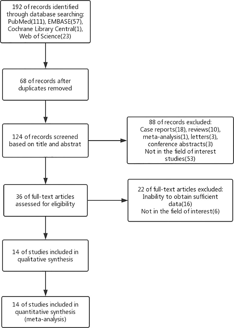
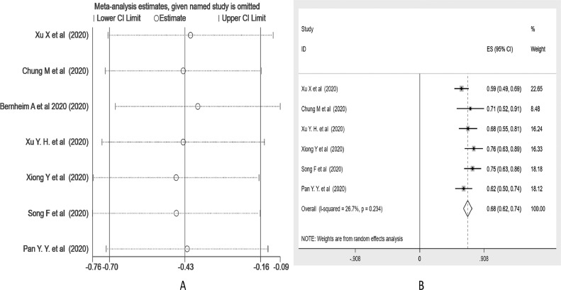
A systematic literature search of PubMed, EMBASE, the Cochrane Library, and Web of Science was performed to identify studies assessing CT features, clinical, and laboratory results of COVID-19 patients. A single-arm meta-analysis was conducted to obtain the pooled prevalence and 95% confidence interval (95% CI).

RESULTS

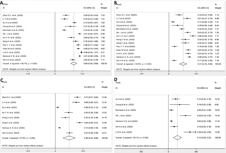
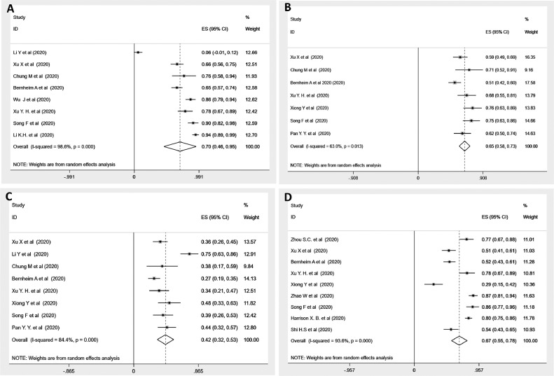
A total of 14 articles (including 1115 patients) based on chest CT images were retrieved. In the lesion patterns on chest CTs, we found that pure ground-glass opacities (GGO) (69%, 95% CI 58-80%), consolidation (47%, 35-60%) and "air bronchogram sign" (46%, 25-66%) were more common than the atypical lesion of "crazy-paving pattern" (15%, 8-22%). With regard to disease extent and involvement, 70% (95% CI 46-95%) of cases showed a location preference for the right lower lobe, 65% (58-73%) of patients presented with ≥3 lobes involvement, and meanwhile, 42% (32-53%) of patients had involvement of all five lobes, while 67% (55-78%) of patients showed a predominant peripheral distribution. An understanding of some important CT features might be helpful for medical surveillance and management. In terms of clinical features, muscle soreness (21%, 95% CI 15-26%) and diarrhea (7%, 4-10%) were minor symptoms compared to fever (80%, 74-87%) and cough (53%, 33-72%).

CONCLUSION

Chest CT manifestations in patients with COVID-19, as well as its main clinical characteristics, might be helpful in disease evolution and management.

摘要

目的

评估胸部 CT 表现的普遍性,根据胸部 CT 图像描述一些显著特征,并介绍 2019 年冠状病毒病(COVID-19)患者的主要临床特征。

材料和方法

对 PubMed、EMBASE、Cochrane 图书馆和 Web of Science 进行系统文献检索,以评估 COVID-19 患者的 CT 特征、临床和实验室结果。进行单臂荟萃分析以获得汇总患病率和 95%置信区间(95%CI)。

结果

共检索到 14 篇基于胸部 CT 图像的研究(包括 1115 例患者)。在胸部 CT 上的病变模式中,我们发现单纯磨玻璃影(GGO)(69%,95%CI 58-80%)、实变(47%,35-60%)和“空气支气管征”(46%,25-66%)比非典型的“铺路石征”(15%,8-22%)更常见。关于疾病程度和受累范围,70%(95%CI 46-95%)的病例显示右下叶位置偏好,65%(58-73%)的患者累及≥3 个肺叶,同时,42%(32-53%)的患者累及所有五个肺叶,而 67%(55-78%)的患者表现为主要的外周分布。了解一些重要的 CT 特征可能有助于医学监测和管理。在临床特征方面,与发热(80%,74-87%)和咳嗽(53%,33-72%)相比,肌肉疼痛(21%,95%CI 15-26%)和腹泻(7%,4-10%)是次要症状。

结论

COVID-19 患者的胸部 CT 表现及其主要临床特征可能有助于疾病的演变和管理。

https://cdn.ncbi.nlm.nih.gov/pmc/blobs/c3a7/7200137/771f772c65c7/gr5_lrg.jpg
https://cdn.ncbi.nlm.nih.gov/pmc/blobs/c3a7/7200137/973ce510add4/gr1_lrg.jpg
https://cdn.ncbi.nlm.nih.gov/pmc/blobs/c3a7/7200137/60f92cd0b904/gr2_lrg.jpg
https://cdn.ncbi.nlm.nih.gov/pmc/blobs/c3a7/7200137/f1d2f82fbd7b/gr3_lrg.jpg
https://cdn.ncbi.nlm.nih.gov/pmc/blobs/c3a7/7200137/c222b0ff3aef/gr4_lrg.jpg
https://cdn.ncbi.nlm.nih.gov/pmc/blobs/c3a7/7200137/771f772c65c7/gr5_lrg.jpg

相似文献

1
CT Manifestations and Clinical Characteristics of 1115 Patients with Coronavirus Disease 2019 (COVID-19): A Systematic Review and Meta-analysis.新冠肺炎患者 1115 例的 CT 表现及临床特征:系统评价和荟萃分析。
Acad Radiol. 2020 Jul;27(7):910-921. doi: 10.1016/j.acra.2020.04.033. Epub 2020 May 5.
2
The chest CT features of coronavirus disease 2019 (COVID-19) in China: a meta-analysis of 19 retrospective studies.中国 2019 年冠状病毒病(COVID-19)的胸部 CT 特征:19 项回顾性研究的荟萃分析。
Virol J. 2020 Oct 21;17(1):159. doi: 10.1186/s12985-020-01432-9.
3
CT imaging features of 34 patients infected with COVID-19.34 例新冠病毒感染患者的 CT 影像学特征。
Clin Imaging. 2020 Dec;68:226-231. doi: 10.1016/j.clinimag.2020.05.016. Epub 2020 May 16.
4
Imaging and clinical features of patients with 2019 novel coronavirus SARS-CoV-2.新型冠状病毒 2019 年 SARS-CoV-2 患者的影像学和临床特征。
Eur J Nucl Med Mol Imaging. 2020 May;47(5):1275-1280. doi: 10.1007/s00259-020-04735-9. Epub 2020 Feb 28.
5
Early Clinical and CT Manifestations of Coronavirus Disease 2019 (COVID-19) Pneumonia.新型冠状病毒肺炎(COVID-19)肺炎的早期临床和 CT 表现。
AJR Am J Roentgenol. 2020 Aug;215(2):338-343. doi: 10.2214/AJR.20.22961. Epub 2020 Mar 17.
6
Spectrum of chest computed tomographic (CT) findings in coronavirus disease-19 (COVID-19) patients in India.印度 COVID-19 患者的胸部计算机断层扫描(CT)表现谱。
Eur J Radiol. 2020 Aug;129:109147. doi: 10.1016/j.ejrad.2020.109147. Epub 2020 Jun 24.
7
Chest CT Findings in Patients With Coronavirus Disease 2019 and Its Relationship With Clinical Features.新型冠状病毒肺炎患者的胸部 CT 表现及其与临床特征的关系。
Invest Radiol. 2020 May;55(5):257-261. doi: 10.1097/RLI.0000000000000670.
8
CT imaging features of 4121 patients with COVID-19: A meta-analysis.4121 例 COVID-19 患者的 CT 影像学特征:一项荟萃分析。
J Med Virol. 2020 Jul;92(7):891-902. doi: 10.1002/jmv.25910. Epub 2020 Apr 29.
9
Analysis of clinical features and imaging signs of COVID-19 with the assistance of artificial intelligence.人工智能辅助分析 COVID-19 的临床特征和影像征象。
Eur Rev Med Pharmacol Sci. 2020 Aug;24(15):8210-8218. doi: 10.26355/eurrev_202008_22510.
10
A Comparison of Clinical and Chest CT Findings in Patients With Influenza A (H1N1) Virus Infection and Coronavirus Disease (COVID-19).甲型流感病毒(H1N1)感染与新型冠状病毒肺炎(COVID-19)患者的临床和胸部 CT 表现比较。
AJR Am J Roentgenol. 2020 Nov;215(5):1065-1071. doi: 10.2214/AJR.20.23214. Epub 2020 May 26.

引用本文的文献

1
The difference of variation types between late-onset multiple acyl-CoA dehydrogenase deficiency patients carrying biallelic and single heterozygous variations in ETFDH: a systematic review and meta-analysis.携带ETFDH双等位基因和单杂合变异的迟发性多种酰基辅酶A脱氢酶缺乏症患者变异类型的差异:一项系统评价和荟萃分析。
Orphanet J Rare Dis. 2025 Jun 18;20(1):310. doi: 10.1186/s13023-025-03845-7.
2
Factors of progression to severity and death in COVID-19 patients at two health care sites in Bamako, Mali.马里巴马科两个医疗点的新冠肺炎患者病情进展至重症和死亡的相关因素。
BMC Infect Dis. 2025 Jan 17;25(1):77. doi: 10.1186/s12879-025-10456-x.
3

本文引用的文献

1
HRCT imaging features in representative imported cases of 2019 novel coronavirus pneumonia.2019新型冠状病毒肺炎典型输入性病例的高分辨率CT成像特征
Precis Clin Med. 2020 Mar;3(1):9-13. doi: 10.1093/pcmedi/pbaa004. Epub 2020 Feb 11.
2
Clinical, laboratory and imaging features of COVID-19: A systematic review and meta-analysis.新型冠状病毒肺炎的临床、实验室和影像学特征:系统评价和荟萃分析。
Travel Med Infect Dis. 2020 Mar-Apr;34:101623. doi: 10.1016/j.tmaid.2020.101623. Epub 2020 Mar 13.
3
Coronavirus Disease 2019 (COVID-19): A Systematic Review of Imaging Findings in 919 Patients.
Early predictors of intensive care unit admission among COVID-19 patients in Qatar.
卡塔尔 COVID-19 患者入住重症监护病房的早期预测指标。
Front Public Health. 2024 Mar 20;12:1278046. doi: 10.3389/fpubh.2024.1278046. eCollection 2024.
4
A Multiclass Radiomics Method-Based WHO Severity Scale for Improving COVID-19 Patient Assessment and Disease Characterization From CT Scans.一种基于多分类放射组学方法的世卫组织严重程度评分标准,用于改善 CT 扫描对 COVID-19 患者的评估和疾病特征描述。
Invest Radiol. 2023 Dec 1;58(12):882-893. doi: 10.1097/RLI.0000000000001005. Epub 2023 Jul 27.
5
Digital tomosynthesis improves chest radiograph accuracy and reduces microbiological false negatives in COVID-19 diagnosis.数字断层合成技术提高了胸部 X 光片的准确率,并降低了 COVID-19 诊断中的微生物假阴性率。
Emerg Radiol. 2023 Aug;30(4):465-474. doi: 10.1007/s10140-023-02153-6. Epub 2023 Jun 26.
6
The Threat of Adversarial Attack on a COVID-19 CT Image-Based Deep Learning System.基于新冠肺炎CT图像的深度学习系统面临对抗攻击的威胁。
Bioengineering (Basel). 2023 Feb 2;10(2):194. doi: 10.3390/bioengineering10020194.
7
COVID19 biomarkers: What did we learn from systematic reviews?COVID19 生物标志物:系统评价给我们带来了哪些启示?
Front Cell Infect Microbiol. 2022 Dec 13;12:1038908. doi: 10.3389/fcimb.2022.1038908. eCollection 2022.
8
Evaluation of chest CT-scan appearances of COVID-19 according to RSNA classification system.根据RSNA分类系统评估新型冠状病毒肺炎的胸部CT扫描表现。
J Family Med Prim Care. 2022 Aug;11(8):4410-4416. doi: 10.4103/jfmpc.jfmpc_8_22. Epub 2022 Aug 30.
9
Rapid artificial intelligence solutions in a pandemic-The COVID-19-20 Lung CT Lesion Segmentation Challenge.大流行中的快速人工智能解决方案——COVID-19-20 肺 CT 病变分割挑战赛。
Med Image Anal. 2022 Nov;82:102605. doi: 10.1016/j.media.2022.102605. Epub 2022 Sep 6.
10
Clinical indicators, nursing diagnoses, and mortality risk in critically ill patients with COVID-19: a retrospective cohort.新冠肺炎危重症患者的临床指标、护理诊断和死亡风险:一项回顾性队列研究。
Rev Esc Enferm USP. 2022 Jul 1;56:e20210568. doi: 10.1590/1980-220X-REEUSP-2021-0568en. eCollection 2022.
新型冠状病毒病 2019(COVID-19):919 例患者影像学表现的系统评价。
AJR Am J Roentgenol. 2020 Jul;215(1):87-93. doi: 10.2214/AJR.20.23034. Epub 2020 Mar 14.
4
COVID-19 patients' clinical characteristics, discharge rate, and fatality rate of meta-analysis.COVID-19 患者的临床特征、出院率和死亡率的荟萃分析。
J Med Virol. 2020 Jun;92(6):577-583. doi: 10.1002/jmv.25757. Epub 2020 Mar 23.
5
Performance of Radiologists in Differentiating COVID-19 from Non-COVID-19 Viral Pneumonia at Chest CT.放射科医生在胸部 CT 鉴别 COVID-19 与非 COVID-19 病毒性肺炎中的表现。
Radiology. 2020 Aug;296(2):E46-E54. doi: 10.1148/radiol.2020200823. Epub 2020 Mar 10.
6
Clinical and High-Resolution CT Features of the COVID-19 Infection: Comparison of the Initial and Follow-up Changes.COVID-19 感染的临床和高分辨率 CT 特征:初始与随访变化比较。
Invest Radiol. 2020 Jun;55(6):332-339. doi: 10.1097/RLI.0000000000000674.
7
CT Features of Coronavirus Disease 2019 (COVID-19) Pneumonia in 62 Patients in Wuhan, China.中国武汉 62 例 2019 年冠状病毒病(COVID-19)肺炎的 CT 特征。
AJR Am J Roentgenol. 2020 Jun;214(6):1287-1294. doi: 10.2214/AJR.20.22975. Epub 2020 Mar 5.
8
Coronavirus Disease 2019 (COVID-19): Role of Chest CT in Diagnosis and Management.新型冠状病毒病 2019(COVID-19):胸部 CT 在诊断和管理中的作用。
AJR Am J Roentgenol. 2020 Jun;214(6):1280-1286. doi: 10.2214/AJR.20.22954. Epub 2020 Mar 4.
9
Relation Between Chest CT Findings and Clinical Conditions of Coronavirus Disease (COVID-19) Pneumonia: A Multicenter Study.新型冠状病毒肺炎的胸部 CT 表现与临床特征的关系:一项多中心研究。
AJR Am J Roentgenol. 2020 May;214(5):1072-1077. doi: 10.2214/AJR.20.22976. Epub 2020 Mar 3.
10
The Clinical and Chest CT Features Associated With Severe and Critical COVID-19 Pneumonia.与严重和危重新冠肺炎相关的临床和胸部 CT 特征。
Invest Radiol. 2020 Jun;55(6):327-331. doi: 10.1097/RLI.0000000000000672.