• 文献检索
  • 文档翻译
  • 深度研究
  • 学术资讯
  • Suppr Zotero 插件Zotero 插件
  • 邀请有礼
  • 套餐&价格
  • 历史记录
应用&插件
Suppr Zotero 插件Zotero 插件浏览器插件Mac 客户端Windows 客户端微信小程序
定价
高级版会员购买积分包购买API积分包
服务
文献检索文档翻译深度研究API 文档MCP 服务
关于我们
关于 Suppr公司介绍联系我们用户协议隐私条款
关注我们

Suppr 超能文献

核心技术专利:CN118964589B侵权必究
粤ICP备2023148730 号-1Suppr @ 2026

文献检索

告别复杂PubMed语法,用中文像聊天一样搜索,搜遍4000万医学文献。AI智能推荐,让科研检索更轻松。

立即免费搜索

文件翻译

保留排版,准确专业,支持PDF/Word/PPT等文件格式,支持 12+语言互译。

免费翻译文档

深度研究

AI帮你快速写综述,25分钟生成高质量综述,智能提取关键信息,辅助科研写作。

立即免费体验

新型冠状病毒肺炎感染的胸部影像学表现。

Chest Imaging Appearance of COVID-19 Infection.

作者信息

Kong Weifang, Agarwal Prachi P

机构信息

Department of Radiology, Sichuan Academy of Medical Science, Sichuan Provincial People's Hospital, Chengdu, China (W.K.); and Division of Cardiothoracic Radiology, Department of Radiology, University of Michigan Health System, 1500 E Medical Center Dr, CVC 5383, Ann Arbor, MI 48109 (P.P.A.).

出版信息

Radiol Cardiothorac Imaging. 2020 Feb 13;2(1):e200028. doi: 10.1148/ryct.2020200028. eCollection 2020 Feb.

DOI:10.1148/ryct.2020200028
PMID:33778544
原文链接:https://pmc.ncbi.nlm.nih.gov/articles/PMC7233424/
Abstract

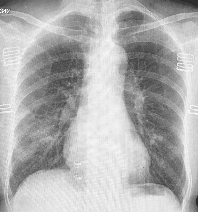
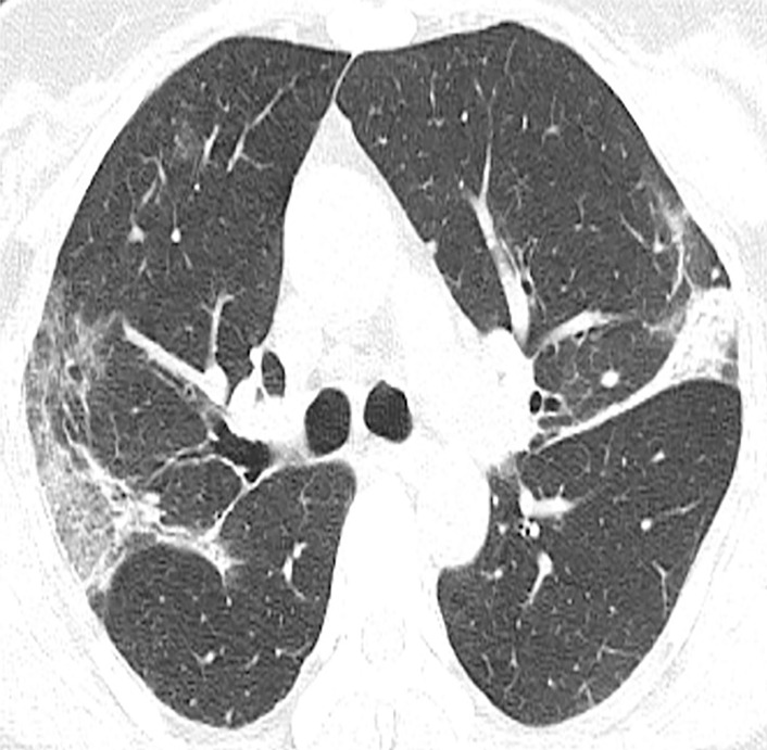
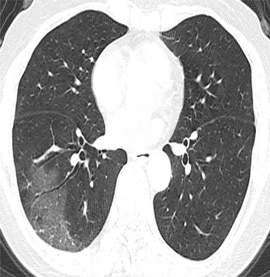
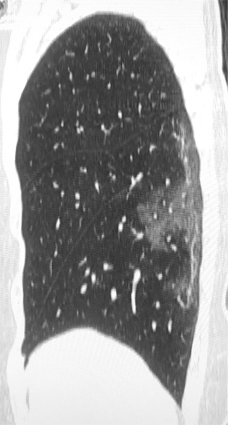
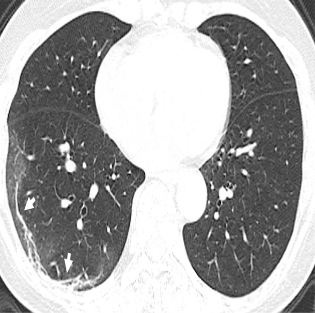
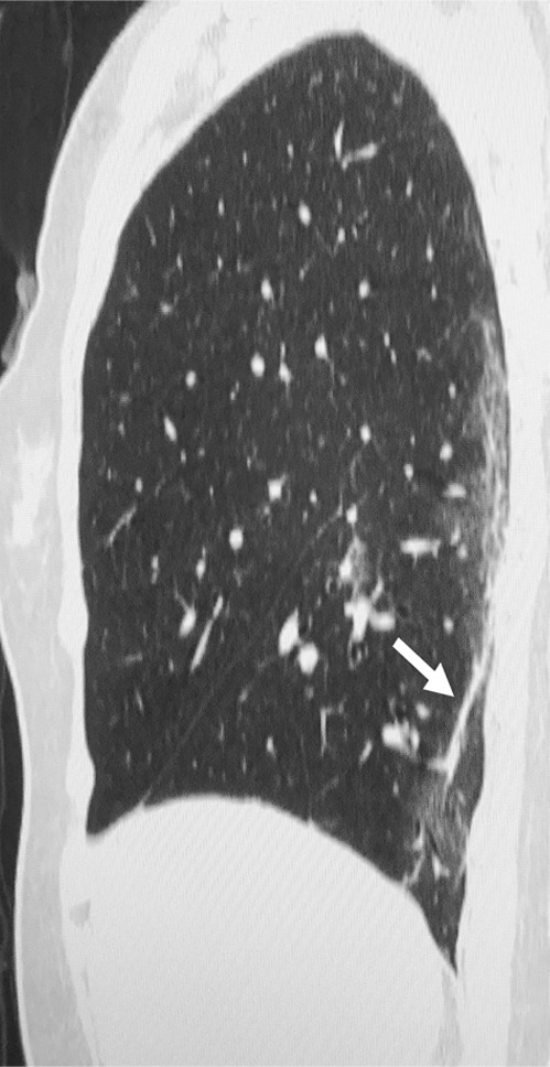
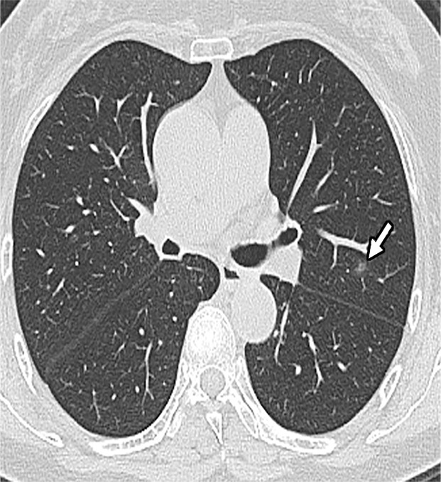
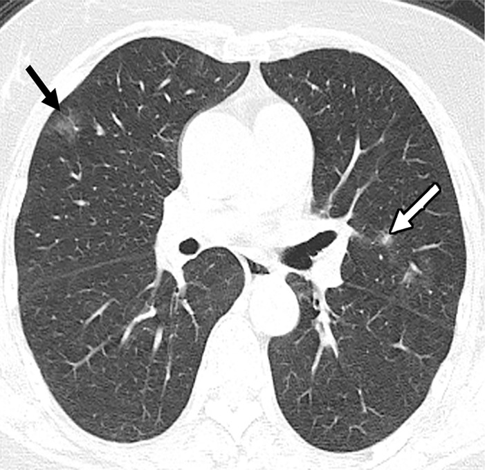
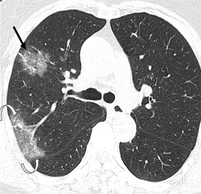
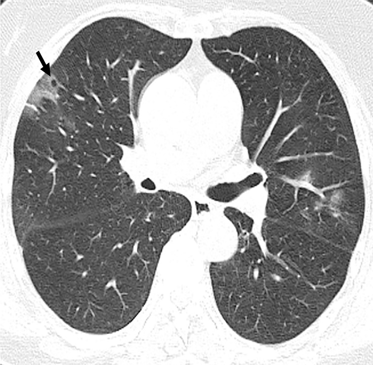
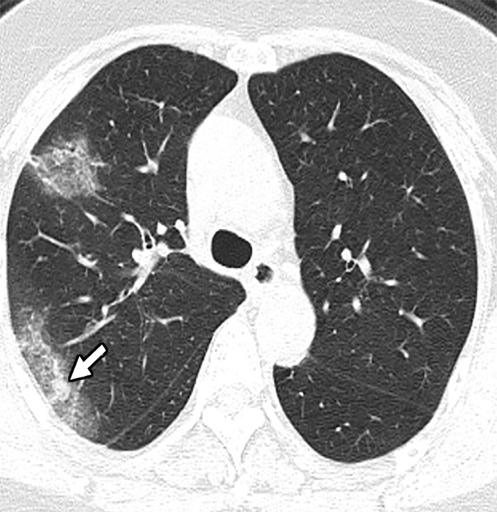
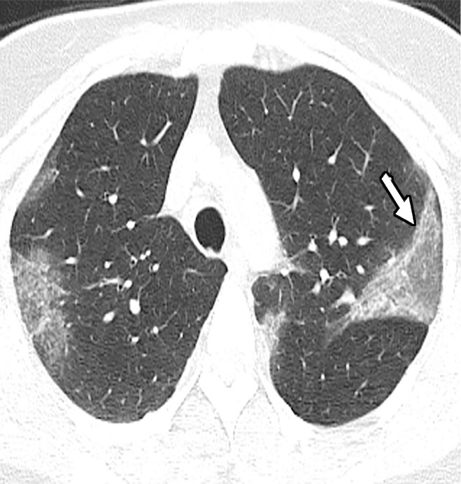
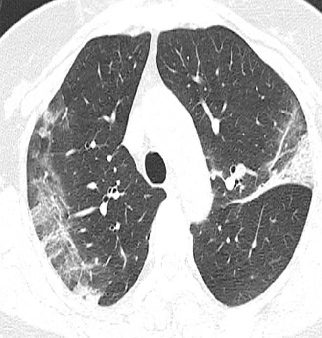
Coronavirus disease 2019 (COVID-19) (previously known as novel coronavirus [2019-nCoV]), first reported in China, has now been declared a global health emergency by the World Health Organization. As confirmed cases are being reported in several countries from all over the world, it becomes important for all radiologists to be aware of the imaging spectrum of the disease and contribute to effective surveillance and response measures. © RSNA, 2020 See editorial by Kay and Abbara in this issue.

摘要

2019冠状病毒病(COVID-19)(先前称为新型冠状病毒[2019-nCoV])首次在中国报告,现已被世界卫生组织宣布为全球卫生突发事件。随着全球多个国家报告确诊病例,所有放射科医生了解该疾病的影像学表现并为有效的监测和应对措施做出贡献变得至关重要。©RSNA,2020 见本期Kay和Abbara的社论。

https://cdn.ncbi.nlm.nih.gov/pmc/blobs/a0ea/7977968/36badffe19cd/ryct.2020200028.fig3c.jpg
https://cdn.ncbi.nlm.nih.gov/pmc/blobs/a0ea/7977968/50fd75da6001/ryct.2020200028.fig1a.jpg
https://cdn.ncbi.nlm.nih.gov/pmc/blobs/a0ea/7977968/ab667727621f/ryct.2020200028.fig1b.jpg
https://cdn.ncbi.nlm.nih.gov/pmc/blobs/a0ea/7977968/765aa5c171dd/ryct.2020200028.fig1c.jpg
https://cdn.ncbi.nlm.nih.gov/pmc/blobs/a0ea/7977968/b854960a8a52/ryct.2020200028.fig1d.jpg
https://cdn.ncbi.nlm.nih.gov/pmc/blobs/a0ea/7977968/f2b055a986ad/ryct.2020200028.fig1e.jpg
https://cdn.ncbi.nlm.nih.gov/pmc/blobs/a0ea/7977968/3fc84694ca23/ryct.2020200028.fig2a.jpg
https://cdn.ncbi.nlm.nih.gov/pmc/blobs/a0ea/7977968/641018487b0b/ryct.2020200028.fig2b.jpg
https://cdn.ncbi.nlm.nih.gov/pmc/blobs/a0ea/7977968/5a517b869660/ryct.2020200028.fig2c.jpg
https://cdn.ncbi.nlm.nih.gov/pmc/blobs/a0ea/7977968/37d1eb2f4adb/ryct.2020200028.fig2d.jpg
https://cdn.ncbi.nlm.nih.gov/pmc/blobs/a0ea/7977968/6bcacaf8ac45/ryct.2020200028.fig2e.jpg
https://cdn.ncbi.nlm.nih.gov/pmc/blobs/a0ea/7977968/dc91c2344ddd/ryct.2020200028.fig3a.jpg
https://cdn.ncbi.nlm.nih.gov/pmc/blobs/a0ea/7977968/ec8863a0fb18/ryct.2020200028.fig3b.jpg
https://cdn.ncbi.nlm.nih.gov/pmc/blobs/a0ea/7977968/36badffe19cd/ryct.2020200028.fig3c.jpg

相似文献

1
Chest Imaging Appearance of COVID-19 Infection.新型冠状病毒肺炎感染的胸部影像学表现。
Radiol Cardiothorac Imaging. 2020 Feb 13;2(1):e200028. doi: 10.1148/ryct.2020200028. eCollection 2020 Feb.
2
2019 novel coronavirus (2019-nCoV) outbreak: A new challenge.2019 新型冠状病毒(2019-nCoV)爆发:新的挑战。
J Glob Antimicrob Resist. 2020 Jun;21:22-27. doi: 10.1016/j.jgar.2020.02.021. Epub 2020 Mar 7.
3
Detection of 2019-Novel Coronavirus (2019-nCoV) by rRT-PCR at Mymensingh Medical College, Mymensingh, Bangladesh.孟加拉国迈门辛医学院利用逆转录实时荧光定量聚合酶链反应(rRT-PCR)检测2019新型冠状病毒(2019-nCoV)
Mymensingh Med J. 2020 Jul;29(3):589-595.
4
Investigating the cases of novel coronavirus disease (COVID-19) in China using dynamic statistical techniques.运用动态统计技术对中国新型冠状病毒肺炎(COVID-19)病例进行调查。
Heliyon. 2020 Apr;6(4):e03747. doi: 10.1016/j.heliyon.2020.e03747. Epub 2020 Apr 4.
5
Clinical Characterizations and Radiological Findings of COVID-19: A 4 Case Report.新型冠状病毒肺炎的临床特征与影像学表现:4例病例报告
Curr Med Imaging. 2022;18(14):1536-1539. doi: 10.2174/1573405618666220404161324.
6
Transmission routes of SARS-CoV-2 and protective measures in dental clinics during the COVID-19 pandemic.2019年冠状病毒病大流行期间严重急性呼吸综合征冠状病毒2的传播途径及牙科诊所的防护措施。
Am J Dent. 2020 Jun;33(3):129-134.
7
Novel coronavirus 2019-nCoV: prevalence, biological and clinical characteristics comparison with SARS-CoV and MERS-CoV.新型冠状病毒 2019-nCoV:流行情况、与 SARS-CoV 和 MERS-CoV 的生物学和临床特征比较。
Eur Rev Med Pharmacol Sci. 2020 Feb;24(4):2012-2019. doi: 10.26355/eurrev_202002_20379.
8
Emergency Radiology During the COVID-19 Pandemic: The Canadian Association of Radiologists Recommendations for Practice.COVID-19 大流行期间的急诊放射学:加拿大放射学家协会的实践推荐。
Can Assoc Radiol J. 2020 Nov;71(4):425-430. doi: 10.1177/0846537120930344. Epub 2020 May 29.
9
The role of chest imaging in the diagnosis, management, and monitoring of coronavirus disease 2019 (COVID-19).胸部影像学在2019冠状病毒病(COVID-19)诊断、管理及监测中的作用
Insights Imaging. 2021 Nov 2;12(1):155. doi: 10.1186/s13244-021-01096-1.
10
Canadian Society of Thoracic Radiology/Canadian Association of Radiologists Consensus Statement Regarding Chest Imaging in Suspected and Confirmed COVID-19.加拿大胸放射学会/加拿大放射学家协会关于疑似和确诊 COVID-19 胸部成像的共识声明。
Can Assoc Radiol J. 2020 Nov;71(4):470-481. doi: 10.1177/0846537120924606. Epub 2020 May 8.

引用本文的文献

1
Pneumothorax and Pneumomediastinum in SARS-CoV-2 Infection.新型冠状病毒肺炎感染中的气胸和纵隔气肿
Medicina (Kaunas). 2025 Jun 29;61(7):1182. doi: 10.3390/medicina61071182.
2
A comparative cohort study of post-COVID-19 conditions based on physical examination records in China.一项基于中国体格检查记录的新冠后状况比较队列研究。
EBioMedicine. 2025 Feb;112:105549. doi: 10.1016/j.ebiom.2024.105549. Epub 2025 Jan 2.
3
Leveraging compact convolutional transformers for enhanced COVID-19 detection in chest X-rays: a grad-CAM visualization approach.

本文引用的文献

1
Chest CT Findings in 2019 Novel Coronavirus (2019-nCoV) Infections from Wuhan, China: Key Points for the Radiologist.中国武汉2019新型冠状病毒(2019-nCoV)感染的胸部CT表现:放射科医生的要点
Radiology. 2020 Apr;295(1):16-17. doi: 10.1148/radiol.2020200241. Epub 2020 Feb 4.
2
CT Imaging Features of 2019 Novel Coronavirus (2019-nCoV).2019 新型冠状病毒(2019-nCoV)的 CT 影像学特征。
Radiology. 2020 Apr;295(1):202-207. doi: 10.1148/radiol.2020200230. Epub 2020 Feb 4.
3
Epidemiological and clinical characteristics of 99 cases of 2019 novel coronavirus pneumonia in Wuhan, China: a descriptive study.
利用紧凑型卷积变压器增强胸部X光片中的COVID-19检测:一种梯度加权类激活映射可视化方法。
Front Big Data. 2024 Dec 16;7:1489020. doi: 10.3389/fdata.2024.1489020. eCollection 2024.
4
Concatenated CNN-Based Pneumonia Detection Using a Fuzzy-Enhanced Dataset.基于串联 CNN 的肺炎检测,使用模糊增强数据集。
Sensors (Basel). 2024 Oct 21;24(20):6750. doi: 10.3390/s24206750.
5
COVID-19 and Pneumonia detection and web deployment from CT scan and X-ray images using deep learning.利用深度学习从 CT 扫描和 X 光图像中检测 COVID-19 和肺炎并进行网络部署。
PLoS One. 2024 Jul 8;19(7):e0302413. doi: 10.1371/journal.pone.0302413. eCollection 2024.
6
Deep learning assisted COVID-19 detection using full CT-scans.使用全胸部CT扫描的深度学习辅助新冠病毒检测
Internet Things (Amst). 2021 Jun;14:100377. doi: 10.1016/j.iot.2021.100377. Epub 2021 Feb 24.
7
Machine learning for medical imaging-based COVID-19 detection and diagnosis.用于基于医学影像的新冠病毒检测与诊断的机器学习
Int J Intell Syst. 2021 Sep;36(9):5085-5115. doi: 10.1002/int.22504. Epub 2021 May 31.
8
Lung pneumonia severity scoring in chest X-ray images using transformers.基于变换模型的胸部 X 射线图像中肺炎严重程度评分
Med Biol Eng Comput. 2024 Aug;62(8):2389-2407. doi: 10.1007/s11517-024-03066-3. Epub 2024 Apr 9.
9
A Hybrid Deep Learning CNN model for COVID-19 detection from chest X-rays.一种用于从胸部X光片中检测新冠肺炎的混合深度学习卷积神经网络模型。
Heliyon. 2024 Feb 29;10(5):e26938. doi: 10.1016/j.heliyon.2024.e26938. eCollection 2024 Mar 15.
10
PulmoNet: a novel deep learning based pulmonary diseases detection model.PulmoNet:一种新型基于深度学习的肺部疾病检测模型。
BMC Med Imaging. 2024 Feb 28;24(1):51. doi: 10.1186/s12880-024-01227-2.
中国武汉 99 例 2019 年新型冠状病毒肺炎患者的流行病学和临床特征:描述性研究。
Lancet. 2020 Feb 15;395(10223):507-513. doi: 10.1016/S0140-6736(20)30211-7. Epub 2020 Jan 30.
4
CT Imaging of the 2019 Novel Coronavirus (2019-nCoV) Pneumonia.2019新型冠状病毒(2019-nCoV)肺炎的CT影像
Radiology. 2020 Apr;295(1):18. doi: 10.1148/radiol.2020200236. Epub 2020 Jan 31.
5
High-resolution CT findings of severe acute respiratory syndrome at presentation and after admission.严重急性呼吸综合征发病时及入院后的高分辨率CT表现
AJR Am J Roentgenol. 2004 Jan;182(1):39-44. doi: 10.2214/ajr.182.1.1820039.