Department of Prosthodontics, University Medical Center Goettingen, Goettingen, Germany.
Department of Prosthetic Dentistry, University Medical Center, Regensburg, Germany.
Clin Oral Investig. 2020 Nov;24(11):3899-3909. doi: 10.1007/s00784-020-03257-w. Epub 2020 Mar 20.
Evidence about modifications of dental luting materials to minimize biological failure at the "marginal gap" between teeth and fixed prosthodontics is scarce. We compared a copper-modified (Co-ZOP) and a conventional zinc oxide phosphate cement (ZOP) in terms of antimicrobial and cytotoxic potentials in vitro and in vivo.
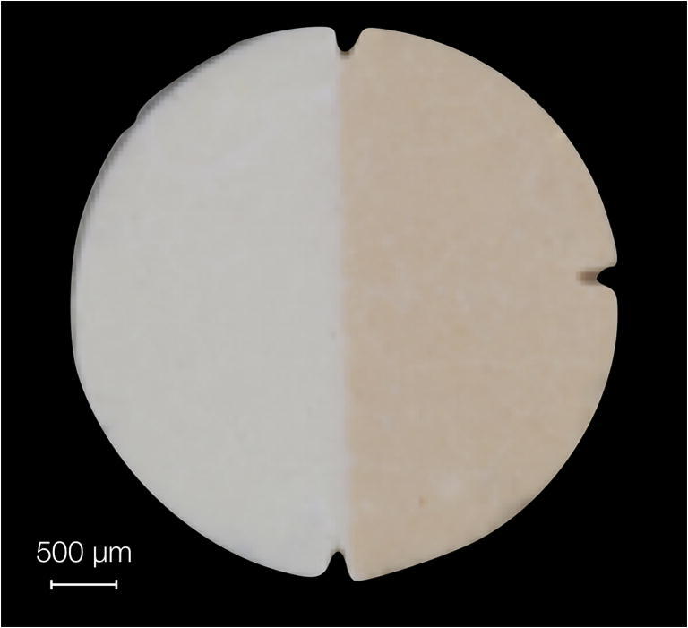
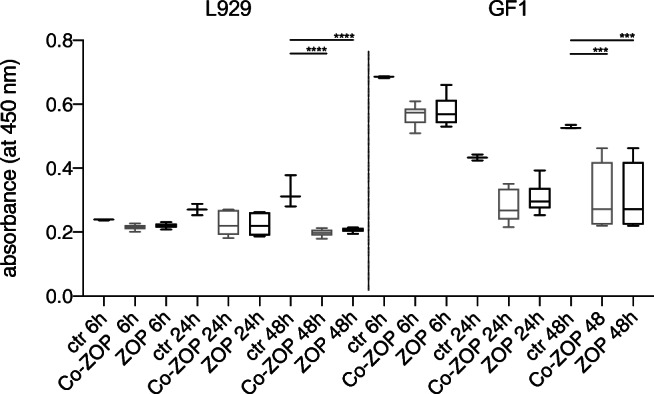
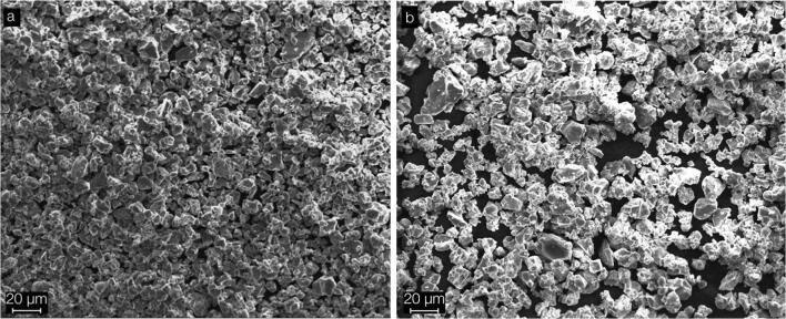
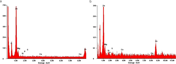
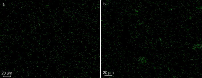
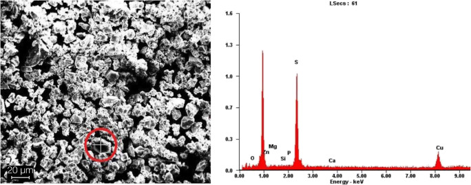
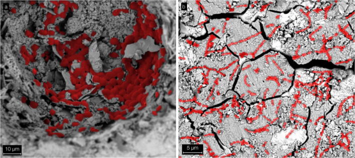
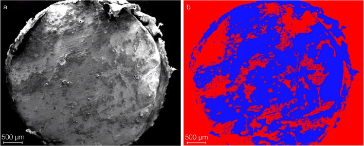
Specimens of ZOP and Co-ZOP were characterized by the mean arithmetic roughness (Ra) and surface free energy (SFE). Powder components were examined using scanning electron microscopy (SEM). Energy-dispersive X-ray spectroscopy (EDX) showed elemental material compositions. In vitro microbial adhesion was shown using SEM, luminescence, and fluorescence assays. CCK-8 assays of mouse fibroblasts (L929) and human gingival fibroblasts (GF-1) were performed after 6, 24, and 48 h of specimen incubation. In vivo, ZOP and Co-ZOP specimens were applied intraorally for 12 h; biofilm accumulation was shown using SEM.
Ra of ZOP and Co-ZOP showed no significant differences; SFE was significantly higher for Co-ZOP. EDX exhibited minor copper radiation for Co-ZOP, none for ZOP. In vitro fungal adhesion to Co-ZOP was significantly higher than to ZOP; in vitro streptococcal adhesion, cytotoxicity, and in vivo biofilm formation were not significantly different.
Co-ZOP showed low surface allocations of copper with no improved antimicrobial properties compared with conventional ZOP in vitro or in vivo.
Antimicrobial effects and low cytotoxicity of biomaterials are important for the clinical outcome. Based on our in vitro and in vivo results, no clinical recommendation can be given for the tested Co-ZOP.
关于牙齿粘固材料改性以最小化牙齿和固定修复体之间“边缘间隙”处生物失败的证据很少。我们比较了一种铜改性(Co-ZOP)和一种传统的氧化锌磷酸酯水泥(ZOP),以评估它们在体外和体内的抗菌和细胞毒性潜力。
通过算术平均粗糙度(Ra)和表面自由能(SFE)来描述 ZOP 和 Co-ZOP 的样本特性。使用扫描电子显微镜(SEM)检查粉末成分。能量色散 X 射线光谱(EDX)显示了元素材料成分。使用 SEM、发光和荧光测定法显示体外微生物粘附。将小鼠成纤维细胞(L929)和人牙龈成纤维细胞(GF-1)的 CCK-8 测定在标本孵育 6、24 和 48 小时后进行。在体内,将 ZOP 和 Co-ZOP 标本应用于口腔内 12 小时;使用 SEM 显示生物膜积累。
ZOP 和 Co-ZOP 的 Ra 没有显著差异;SFE 对 Co-ZOP 显著更高。EDX 显示 Co-ZOP 有少量铜辐射,ZOP 则没有。体外真菌对 Co-ZOP 的粘附明显高于 ZOP;体外链球菌粘附、细胞毒性和体内生物膜形成没有显著差异。
与传统 ZOP 相比,Co-ZOP 显示出较低的铜表面分配,但在体外或体内均未显示出改善的抗菌性能。
生物材料的抗菌效果和低细胞毒性对临床结果很重要。基于我们的体外和体内结果,不能对测试的 Co-ZOP 给出临床建议。