• 文献检索
  • 文档翻译
  • 深度研究
  • 学术资讯
  • Suppr Zotero 插件Zotero 插件
  • 邀请有礼
  • 套餐&价格
  • 历史记录
应用&插件
Suppr Zotero 插件Zotero 插件浏览器插件Mac 客户端Windows 客户端微信小程序
定价
高级版会员购买积分包购买API积分包
服务
文献检索文档翻译深度研究API 文档MCP 服务
关于我们
关于 Suppr公司介绍联系我们用户协议隐私条款
关注我们

Suppr 超能文献

核心技术专利:CN118964589B侵权必究
粤ICP备2023148730 号-1Suppr @ 2026

文献检索

告别复杂PubMed语法,用中文像聊天一样搜索,搜遍4000万医学文献。AI智能推荐,让科研检索更轻松。

立即免费搜索

文件翻译

保留排版,准确专业,支持PDF/Word/PPT等文件格式,支持 12+语言互译。

免费翻译文档

深度研究

AI帮你快速写综述,25分钟生成高质量综述,智能提取关键信息,辅助科研写作。

立即免费体验

人类晚期种植体周围炎骨缺损的组织学特征。

Histological characteristics of advanced peri-implantitis bone defects in humans.

作者信息

Galárraga-Vinueza Maria Elisa, Tangl Stefan, Bianchini Marco, Magini Ricardo, Obreja Karina, Gruber Reinhard, Schwarz Frank

机构信息

Department of Oral Surgery and Implantology, Carolinum, Goethe University, Frankfurt, Germany.

Post-Graduate Program in Implant Dentistry (PPGO), Federal University of Santa Catarina (UFSC), Florianópolis, SC, Brazil.

出版信息

Int J Implant Dent. 2020 Mar 25;6(1):12. doi: 10.1186/s40729-020-00208-8.

DOI:10.1186/s40729-020-00208-8
PMID:32211972
原文链接:https://pmc.ncbi.nlm.nih.gov/articles/PMC7093613/
Abstract

BACKGROUND

Inflammatory osteolysis is the clinical hallmark of peri-implantitis. The morphology of the remaining peri-implant bone and the level of osseointegration, however, remain unknown. Our aim was to characterize advanced peri-implantitis bone defects in humans.

METHODS

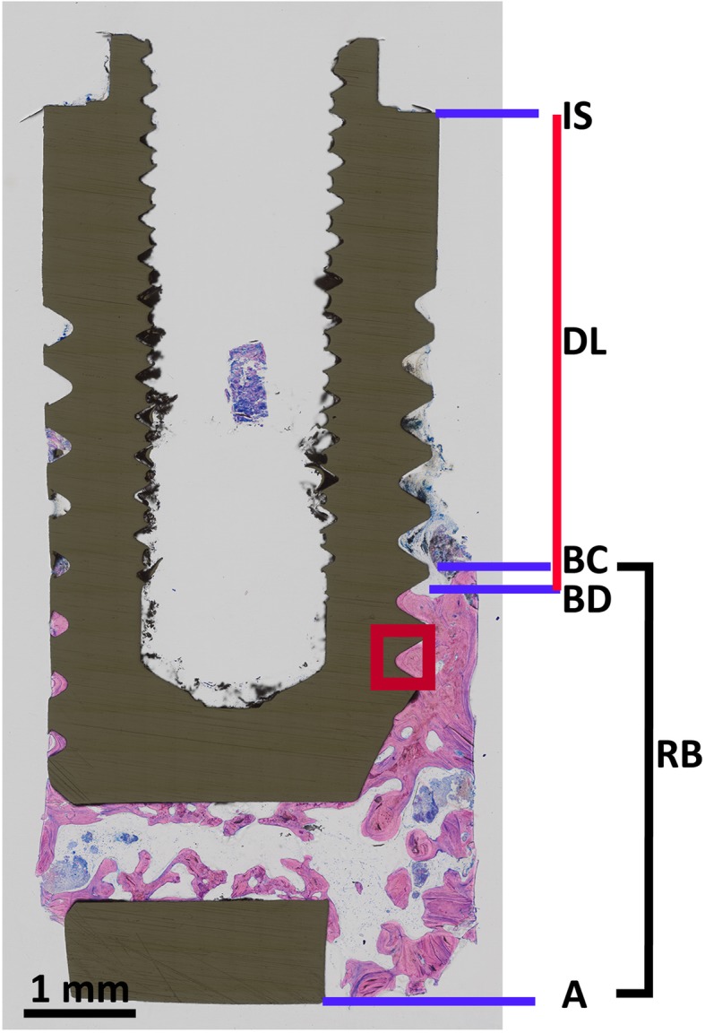
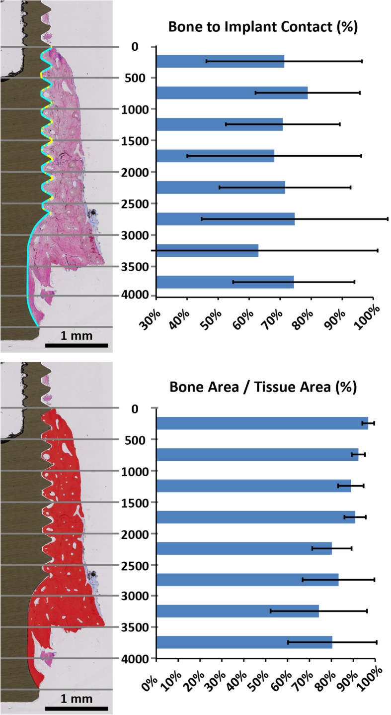
Four patients (3 female and 1 male) were diagnosed with peri-implantitis. A total of 5 implants with machined surfaces and a mean loading time of 12 ± 6 years were removed due to advanced bone loss. The defect extension, the peri-implant bone density (bone area per tissue area in percentage), bone-to-implant contact (%), and the number of filled and empty osteocyte lacunae were calculated based on undecalcified histological specimens.

RESULTS

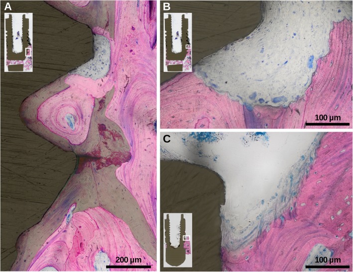
The defect extension was on average 4.2 mm (95% CI 0.8-3.4). Remaining peri-implant bone showed a high density of 85.5% (95% CI 79.1-91.3) and covered in total 74% (95% CI 70.5-77.5) of the implant surface. Filled and empty osteocyte lacunae density was on average 191 and 165/mm (95% CI 132-251; 103-225), respectively. Histology further revealed signs of ongoing bone formation and resorption.

CONCLUSION

There are signs that suggest that once the original cortical bone is lost due to peri-implantitis, the remaining apical trabecular bone is reinforced and transformed into cortical bone that might take over the functional load.

摘要

背景

炎性骨溶解是种植体周围炎的临床特征。然而,剩余种植体周围骨的形态以及骨结合水平仍不清楚。我们的目的是对人类晚期种植体周围炎骨缺损进行特征描述。

方法

4例患者(3例女性,1例男性)被诊断为种植体周围炎。由于严重骨吸收,共取出5枚表面经过机械加工、平均植入时间为12±6年的种植体。基于未脱钙组织学标本计算缺损范围、种植体周围骨密度(骨面积占组织面积的百分比)、骨与种植体的接触面积(%)以及填充和空的骨细胞陷窝数量。

结果

缺损范围平均为4.2 mm(95%可信区间0.8 - 3.4)。剩余种植体周围骨显示高密度为85.5%(95%可信区间79.1 -

https://cdn.ncbi.nlm.nih.gov/pmc/blobs/7c60/7093613/8130fd73eabf/40729_2020_208_Fig3_HTML.jpg
https://cdn.ncbi.nlm.nih.gov/pmc/blobs/7c60/7093613/69d44813104d/40729_2020_208_Fig1_HTML.jpg
https://cdn.ncbi.nlm.nih.gov/pmc/blobs/7c60/7093613/4a9a942562a1/40729_2020_208_Fig2_HTML.jpg
https://cdn.ncbi.nlm.nih.gov/pmc/blobs/7c60/7093613/8130fd73eabf/40729_2020_208_Fig3_HTML.jpg
https://cdn.ncbi.nlm.nih.gov/pmc/blobs/7c60/7093613/69d44813104d/40729_2020_208_Fig1_HTML.jpg
https://cdn.ncbi.nlm.nih.gov/pmc/blobs/7c60/7093613/4a9a942562a1/40729_2020_208_Fig2_HTML.jpg
https://cdn.ncbi.nlm.nih.gov/pmc/blobs/7c60/7093613/8130fd73eabf/40729_2020_208_Fig3_HTML.jpg

相似文献

1
Histological characteristics of advanced peri-implantitis bone defects in humans.人类晚期种植体周围炎骨缺损的组织学特征。
Int J Implant Dent. 2020 Mar 25;6(1):12. doi: 10.1186/s40729-020-00208-8.
2
Morphology and severity of peri-implantitis bone defects.种植体周围炎骨缺损的形态和严重程度。
Clin Implant Dent Relat Res. 2019 Aug;21(4):635-643. doi: 10.1111/cid.12791. Epub 2019 May 14.
3
Re-osseointegration following reconstructive surgical therapy of experimental peri-implantitis. A pre-clinical in vivo study.实验性种植体周围炎重建性外科治疗后的再骨整合:一项临床前体内研究。
Clin Oral Implants Res. 2019 May;30(5):447-456. doi: 10.1111/clr.13430. Epub 2019 May 3.
4
Long time follow up of implant therapy and treatment of peri-implantitis.种植体治疗及种植体周围炎治疗的长期随访
Swed Dent J Suppl. 2007(188):7-66.
5
Three-Year Evaluation of the Influence of Implant Surfaces on Implant Failure and Peri-implantitis: A Double-Blind Randomized Controlled Trial with Split-Mouth Design.种植体表面对种植体失败和种植体周围炎影响的三年评估:一项采用双侧随机对照试验和分口设计的临床试验。
Int J Oral Maxillofac Implants. 2021 Mar-Apr;36(2):e23-e30. doi: 10.11607/jomi.8538.
6
Features of peri-implantitis associated with luting cement extrusion: a histopathologic report in humans.与粘结水门汀挤出相关的种植体周围炎特征:一份人体组织病理学报告
Quintessence Int. 2020;51(5):398-404. doi: 10.3290/j.qi.a44369.
7
The adjunctive effect of rhBMP-2 on the regeneration of peri-implant bone defects after experimental peri-implantitis.rhBMP-2 对实验性种植体周围炎后种植体周围骨缺损再生的辅助作用。
Clin Oral Implants Res. 2019 Dec;30(12):1209-1219. doi: 10.1111/clr.13534. Epub 2019 Sep 24.
8
Coated vs uncoated implants: bone defect configurations after progressive peri-implantitis in dogs.涂层与未涂层种植体:犬种植体周围炎进展后的骨缺损形态
J Oral Implantol. 2014 Dec;40(6):661-9. doi: 10.1563/AAID-JOI-D-12-00089.
9
Re-osseointegration after treatment of peri-implantitis at different implant surfaces. An experimental study in the dog.不同种植体表面种植体周围炎治疗后的再骨结合。犬类实验研究。
Clin Oral Implants Res. 2001 Dec;12(6):595-603. doi: 10.1034/j.1600-0501.2001.120607.x.
10
Role of toll-like receptor 2 in inflammation and alveolar bone loss in experimental peri-implantitis versus periodontitis.Toll 样受体 2 在实验性种植体周围炎与牙周炎中的炎症和牙槽骨丧失中的作用。
J Periodontal Res. 2018 Feb;53(1):98-106. doi: 10.1111/jre.12492. Epub 2017 Sep 5.

引用本文的文献

1
Prevalence, incidence, systemic, behavioral, and patient-related risk factors and indicators for peri-implant diseases: An AO/AAP  systematic review and meta-analysis.种植体周围疾病的患病率、发病率、全身、行为及患者相关危险因素和指标:一项AO/AAP系统评价与Meta分析
J Periodontol. 2025 Jun;96(6):587-633. doi: 10.1002/JPER.24-0154. Epub 2025 Jun 9.
2
CBCT Evaluation of Maxillary Incisive Canal Characteristics Among Population in Regard to Possibility of Implant Cortical Anchorage-A Multicenter Study.CBCT评估上颌切牙管特征在种植体皮质骨锚固可能性方面的人群差异——一项多中心研究
Dent J (Basel). 2025 May 14;13(5):211. doi: 10.3390/dj13050211.
3

本文引用的文献

1
Morphology and severity of peri-implantitis bone defects.种植体周围炎骨缺损的形态和严重程度。
Clin Implant Dent Relat Res. 2019 Aug;21(4):635-643. doi: 10.1111/cid.12791. Epub 2019 May 14.
2
Two to six-year disease resolution and marginal bone stability rates of a modified resective-implantoplasty therapy in 32 peri-implantitis cases.32 例种植体周围炎经改良切除-种植体再成形治疗后 2 至 6 年的疾病缓解率和边缘骨稳定性。
Clin Implant Dent Relat Res. 2019 Aug;21(4):758-765. doi: 10.1111/cid.12773. Epub 2019 Apr 15.
3
Osteoimmunology: Inflammatory osteolysis and regeneration of the alveolar bone.
Histomorphometric Analysis of Osseointegrated Intraosseous Dental Implants Using Undecalcified Specimens: A Scoping Review.
使用未脱钙标本对骨结合型骨内牙种植体进行组织形态计量学分析:一项综述。
Biomimetics (Basel). 2024 Nov 3;9(11):672. doi: 10.3390/biomimetics9110672.
4
Loss of Fine Motor Dexterity and Reduced Oral Hygiene 15 Years After Diagnosis of Multifocal Motor Neuropathy with Screw-Retained Implant-Supported Rehabilitation: Literature Review and Case Report.多灶运动神经病诊断 15 年后丧失精细运动灵巧度和口腔卫生状况下降:文献回顾和病例报告。
Oral Health Prev Dent. 2024 Nov 18;22:583-594. doi: 10.3290/j.ohpd.b5828022.
5
Challenges and Pitfalls of Research Designs Involving Magnesium-Based Biomaterials: An Overview.涉及镁基生物材料的研究设计的挑战和陷阱:概述。
Int J Mol Sci. 2024 Jun 5;25(11):6242. doi: 10.3390/ijms25116242.
6
Ultrastructural and immunohistochemical evaluation of hyperplastic soft tissues surrounding dental implants in fibular jaws.腓骨下颌骨中种植体周围增生软组织的超微结构和免疫组织化学评估。
Sci Rep. 2024 May 10;14(1):10717. doi: 10.1038/s41598-024-60474-z.
7
Echo-intensity characterization at implant sites and novel diagnostic ultrasonographic markers for peri-implantitis.种植体部位的回声强度特征及种植体周围炎的新型诊断超声标志物。
J Clin Periodontol. 2024 Dec;51(12):1586-1597. doi: 10.1111/jcpe.13976. Epub 2024 Apr 1.
8
Impact of Smoking Habit on Peri-Implant Indicators following Different Therapies: A Systematic Review.不同治疗方法后吸烟习惯对种植体周围指标的影响:一项系统评价
Bioengineering (Basel). 2022 Oct 18;9(10):569. doi: 10.3390/bioengineering9100569.
9
Effects of implant surface mechanical instrumentation methods on peri-implantitis: An study using a circumferential bone defect model.种植体表面机械处理方法对种植体周围炎的影响:一项使用环形骨缺损模型的研究。
J Dent Sci. 2022 Apr;17(2):891-896. doi: 10.1016/j.jds.2021.08.018. Epub 2021 Sep 14.
10
Pilot Study of Use of Nitric Oxide in Monitoring Multiple Dental Foci in Oral Cavity-A Case Report.一氧化氮用于监测口腔多牙病灶的初步研究——病例报告
Healthcare (Basel). 2022 Jan 20;10(2):195. doi: 10.3390/healthcare10020195.
骨免疫学:炎症性骨溶解与牙槽骨再生。
J Clin Periodontol. 2019 Jun;46 Suppl 21:52-69. doi: 10.1111/jcpe.13056.
4
Peri-implant diseases and conditions: Consensus report of workgroup 4 of the 2017 World Workshop on the Classification of Periodontal and Peri-Implant Diseases and Conditions.种植体周围疾病和状况:2017 年牙周病和种植体周围病分类世界研讨会第 4 工作组的共识报告。
J Clin Periodontol. 2018 Jun;45 Suppl 20:S286-S291. doi: 10.1111/jcpe.12957.
5
Peri-implantitis.种植体周围炎。
J Clin Periodontol. 2018 Jun;45 Suppl 20:S246-S266. doi: 10.1111/jcpe.12954.
6
Osteocytes play an important role in experimental periodontitis in healthy and diabetic mice through expression of RANKL.成骨细胞通过表达 RANKL 在健康和糖尿病小鼠的实验性牙周炎中发挥重要作用。
J Clin Periodontol. 2018 Mar;45(3):285-292. doi: 10.1111/jcpe.12851. Epub 2018 Jan 25.
7
Osteocyte Mechanobiology.骨细胞机械生物学。
Curr Osteoporos Rep. 2017 Aug;15(4):318-325. doi: 10.1007/s11914-017-0373-0.
8
Peri-implantitis - onset and pattern of progression.种植体周围炎——发病机制和进展模式。
J Clin Periodontol. 2016 Apr;43(4):383-8. doi: 10.1111/jcpe.12535. Epub 2016 Mar 29.
9
Risk indicators for Peri-implantitis. A cross-sectional study with 916 implants.种植体周围炎的风险指标。一项对916颗种植体的横断面研究。
Clin Oral Implants Res. 2017 Feb;28(2):144-150. doi: 10.1111/clr.12772. Epub 2016 Jan 11.
10
Accuracy of periapical radiography in assessing bone level in implants affected by peri-implantitis: a cross-sectional study.根尖片评估种植体周围炎影响的种植体骨水平的准确性:一项横断面研究。
J Clin Periodontol. 2016 Jan;43(1):85-91. doi: 10.1111/jcpe.12491. Epub 2016 Feb 12.