Deng Fengbin, Mu Changping, Yang Ling, Li Huaqinag, Xiang Xuemei, Li Kang, Yang Qingjun
Radiology Department, Chongqing General Hospital, University of Chinese Academy of Science.
Jane lab, Big Data Research Center, University of Electronic Science and Technology of China, China.
Medicine (Baltimore). 2020 Mar;99(13):e19377. doi: 10.1097/MD.0000000000019377.
MRI findings of carotid plaque components have been studied recently as a tool to predict recurrent ischemic events. We performed a systematic review and meta-analysis to summarize the association of MRI-determined intraplaque hemorrhage, lipid-rich necrotic core, and thinning/rupture of the fibrous cap with recurrent ischemic events.
Electronic search was performed in PUBMED, EMBASE, Cochrane Controlled Register of Trials (CENTRAL) from inception to Oct 30, 2018. We included cohort studies with an average follow-up time of more than 1 month in which intraplaque hemorrhage, lipid-rich necrotic core, or thinning/rupture of the fibrous cap were associated with recurrent ipsilateral stroke or ischemic events. We performed heterogeneity assessment before carrying out meta-analysis. According to the heterogeneity, we selected fixed-effect model for meta-analysis of the included cohort studies.
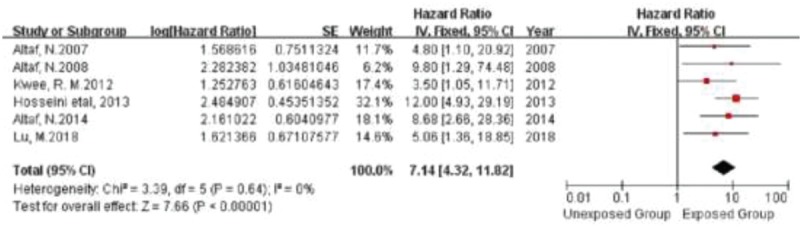
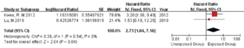
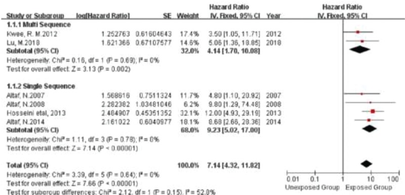
Using a prespecified search strategy, of the 2128 articles, 6 studies with a total number of 621 participants met eligibility for systematic review and meta-analysis. The hazard ratios of intra-plaque hemorrhage, thinning/rupture of the fibrous cap and lipid rich necrotic core as recurrent Stroke/Transient ischemic attack (TIA) were 7.14(95% confidence interval, 4.32 to 11.82), 5.68(95% confidence interval, 2.40 to 13.47), and 2.73(95% confidence interval, 1.04 to 7.16), respectively. No significant heterogeneity was found in the 3 meta-analyses.
The presence of intraplaque hemorrhage, lipid-rich necrotic core, and thinning/rupture of the fibrous cap on MRI of carotid plaque are strong predictors of recurrent stroke events. However, due to the lack of original studies, larger cohort studies are warranted.
近年来,颈动脉斑块成分的磁共振成像(MRI)表现已被作为预测复发性缺血事件的一种工具进行研究。我们进行了一项系统评价和荟萃分析,以总结MRI确定的斑块内出血、富含脂质的坏死核心以及纤维帽变薄/破裂与复发性缺血事件之间的关联。
在PUBMED、EMBASE、Cochrane对照试验注册中心(CENTRAL)进行了从数据库建立至2018年10月30日的电子检索。我们纳入了平均随访时间超过1个月的队列研究,这些研究中斑块内出血、富含脂质的坏死核心或纤维帽变薄/破裂与同侧复发性卒中或缺血事件相关。在进行荟萃分析之前,我们进行了异质性评估。根据异质性情况,我们选择固定效应模型对纳入的队列研究进行荟萃分析。
采用预先设定的检索策略,在2128篇文章中,有6项研究共621名参与者符合系统评价和荟萃分析的纳入标准。斑块内出血、纤维帽变薄/破裂以及富含脂质的坏死核心作为复发性卒中/短暂性脑缺血发作(TIA)的风险比分别为7.14(95%置信区间,4.32至11.82)、5.68(95%置信区间,2.40至13.47)和2.73(95%置信区间,1.04至7.16)。这三项荟萃分析均未发现显著的异质性。
颈动脉斑块MRI上存在斑块内出血、富含脂质的坏死核心以及纤维帽变薄/破裂是复发性卒中事件的有力预测指标。然而,由于缺乏原始研究,有必要开展更大规模的队列研究。