School of Engineering, University of Warwick, Coventry, UK.
School of Pharmacy and Bioengineering, Keele University, Stoke-on-Trent, UK.
Angew Chem Int Ed Engl. 2020 Jul 13;59(29):11984-11991. doi: 10.1002/anie.202000239. Epub 2020 May 14.
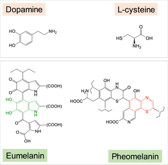
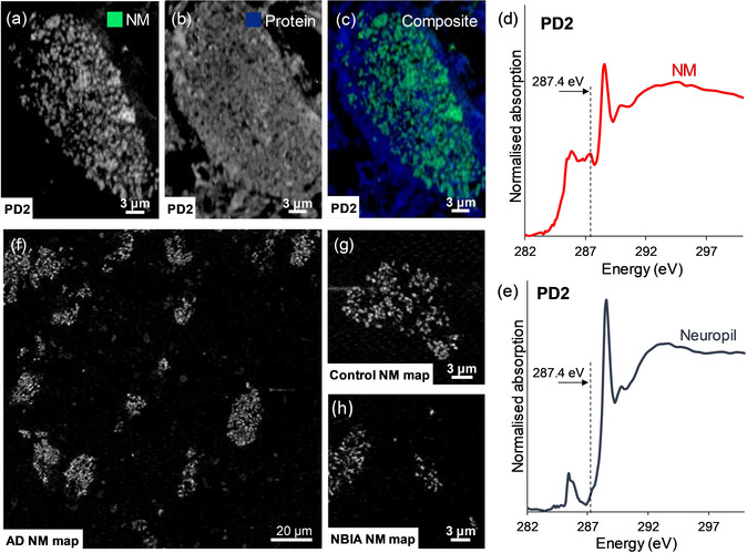
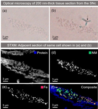
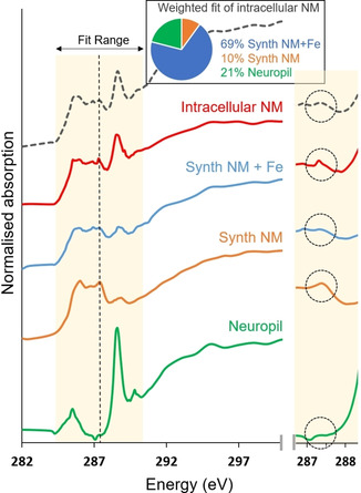
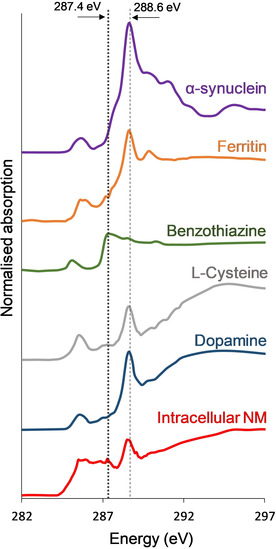
A hallmark of Parkinson's disease is the death of neuromelanin-pigmented neurons, but the role of neuromelanin is unclear. The in situ characterization of neuromelanin remains dependent on detectable pigmentation, rather than direct quantification of neuromelanin. We show that direct, label-free nanoscale visualization of neuromelanin and associated metal ions in human brain tissue can be achieved using synchrotron scanning transmission x-ray microscopy (STXM), through a characteristic feature in the neuromelanin x-ray absorption spectrum at 287.4 eV that is also present in iron-free and iron-laden synthetic neuromelanin. This is confirmed in consecutive brain sections by correlating STXM neuromelanin imaging with silver nitrate-stained neuromelanin. Analysis suggests that the 1s-σ* (C-S) transition in benzothiazine groups accounts for this feature. This method illustrates the wider potential of STXM as a label-free spectromicroscopy technique applicable to both organic and inorganic materials.
帕金森病的一个标志是神经黑色素着色神经元的死亡,但神经黑色素的作用尚不清楚。神经黑色素的原位特征仍然依赖于可检测的色素沉着,而不是对神经黑色素的直接定量。我们表明,使用同步加速器扫描透射 X 射线显微镜(STXM)可以直接、无标记地对人脑组织中的神经黑色素和相关金属离子进行纳米级可视化,这是通过神经黑色素 X 射线吸收光谱在 287.4 eV 处的特征来实现的,该特征也存在于无铁和含铁的合成神经黑色素中。通过将 STXM 神经黑色素成像与硝酸银染色的神经黑色素进行关联,在连续的脑组织切片中得到了证实。分析表明,苯并噻嗪基团的 1s-σ*(C-S)跃迁解释了这一特征。该方法说明了 STXM 作为一种适用于有机和无机材料的无标记光谱显微镜技术的更广泛潜力。