Department of Burns, Nanfang Hospital, Southern Medical University, Jingxi Street, Baiyun District, Guangzhou, 510515, People's Republic of China.
Guangdong Engineering Research Center of Implantable Medical Polymer, Shenzhen Lando Biomaterials Co., Ltd., Shenzhen, 518107, People's Republic of China.
Stem Cell Res Ther. 2020 Mar 31;11(1):141. doi: 10.1186/s13287-020-01645-3.
Three-dimensional (3D) cultivation with biomaterials was proposed to facilitate stem cell epithelial differentiation for wound healing. However, whether human adipose-derived stem cells (hASCs) on collagen sponge scaffold (CSS) better differentiate to keratinocytes remains unclear.
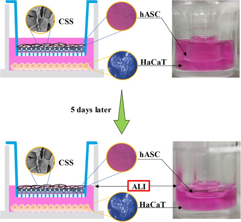
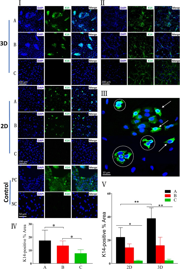
3D cultivation with CSS on hASC epidermal differentiation co-cultured with HaCaT cells at air-liquid interface (ALI) was compared with two-dimensional (2D) form and cultivation without "co-culture" or "ALI." Cellular morphology, cell adhesion, and growth condition were evaluated, followed by the protein and gene expression of keratin 14 (K14, keratinocyte specific marker).
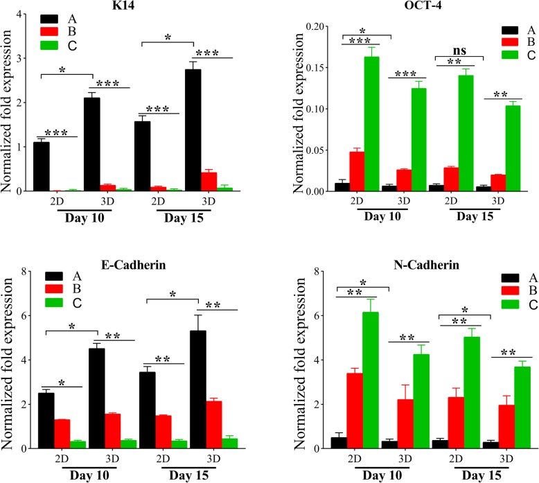
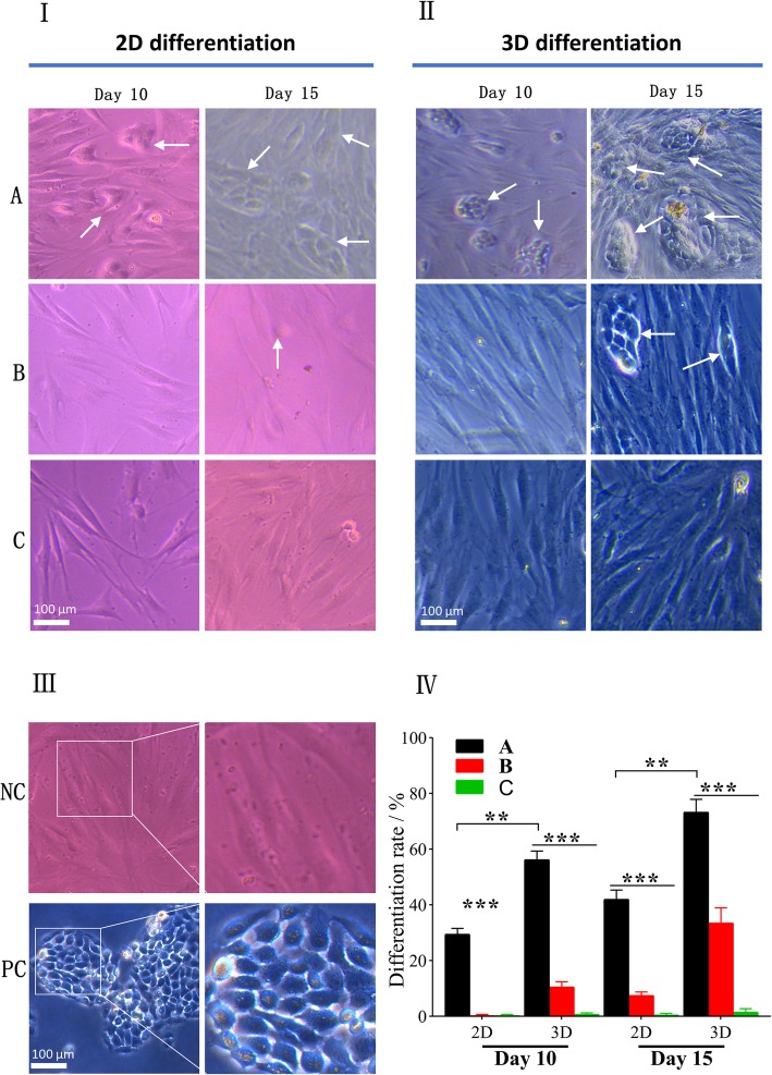
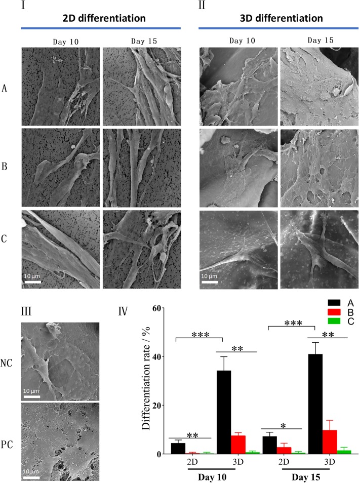
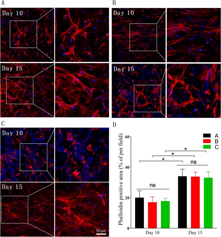
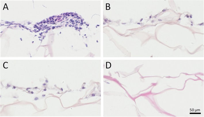
Typical cobblestone morphology of keratinocytes was remarkably observed in co-cultured hASCs at ALI, but those seeded on the CSS exhibited more keratinocyte-like cells under an invert microscope and scanning electron microscope. Desired cell adhesion and proliferation were confirmed in 3D differentiation groups by rhodamine-labeled phalloidin staining, consistent with H&E staining. Compared with those cultured in 2D culture system or without "ALI," immunofluorescence staining and gene expression analysis revealed hASCs co-cultured over CSS expressed K14 at higher levels at day 15.
CSS is positive to promote epithelial differentiation of hASCs, which will foster a deeper understanding of artificial dermis in skin wound healing and regeneration.
三维(3D)培养与生物材料被提出,以促进干细胞上皮分化,用于伤口愈合。然而,人脂肪来源干细胞(hASCs)在胶原海绵支架(CSS)上是否更有利于向角质细胞分化尚不清楚。
在气液界面(ALI)上与 HaCaT 细胞共培养的 hASC 表皮分化的 3D 培养与二维(2D)形式和无“共培养”或“ALI”的培养进行比较。评估细胞形态、细胞黏附和生长情况,然后检测角蛋白 14(K14,角质细胞特异性标志物)的蛋白和基因表达。
在共培养的 ALI 中的 hASCs 中,可明显观察到典型的鹅卵石状角质细胞形态,而在 CSS 上接种的细胞在倒置显微镜和扫描电子显微镜下表现出更多的角质细胞样细胞。通过罗丹明标记鬼笔环肽染色证实 3D 分化组中所需的细胞黏附和增殖,与 H&E 染色一致。与在 2D 培养系统中或没有“ALI”培养的细胞相比,免疫荧光染色和基因表达分析显示,共培养在 CSS 上的 hASCs 在第 15 天表达更高水平的 K14。
CSS 有利于促进 hASCs 的上皮分化,这将促进对皮肤伤口愈合和再生中人工真皮的更深入理解。