Department of Radiology, Jiangmen Central Hospital, Affiliated Jiangmen Hospital of Sun Yat-Sen University, No. 23 Haibang Street, Jiangmen, 529000, Guangdong, China.
Department of Gastrointestinal Surgery, Jiangmen Central Hospital, Affiliated Jiangmen Hospital of Sun Yat-Sen University, Jiangmen, Guangdong, China.
BMC Cancer. 2020 Apr 3;20(1):274. doi: 10.1186/s12885-020-6712-z.
Lymphovascular invasion (LVI) has never been revealed by preoperative scans. It is necessary to use digital mammography in predicting LVI in patients with breast cancer preoperatively.
Overall 122 cases of invasive ductal carcinoma diagnosed between May 2017 and September 2018 were enrolled and assigned into the LVI positive group (n = 42) and the LVI negative group (n = 80). Independent t-test and χ2 test were performed.
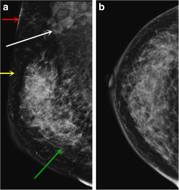
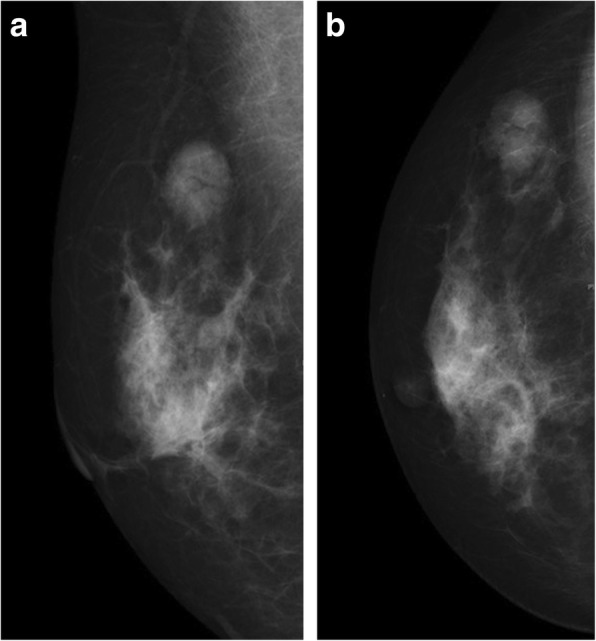
Difference in Ki-67 between the two groups was statistically significant (P = 0.012). Differences in interstitial edema (P = 0.013) and skin thickening (P = 0.000) were statistically significant between the two groups. Multiple factor analysis showed that there were three independent risk factors for LVI: interstitial edema (odds ratio [OR] = 12.610; 95% confidence interval [CI]: 1.061-149.922; P = 0.045), blurring of subcutaneous fat (OR = 0.081; 95% CI: 0.012-0.645; P = 0.017) and skin thickening (OR = 9.041; 95% CI: 2.553-32.022; P = 0.001).
Interstitial edema, blurring of subcutaneous fat, and skin thickening are independent risk factors for LVI. The specificity of LVI prediction is as high as 98.8% when the three are used together.
术前扫描从未显示过淋巴血管侵犯(LVI)。有必要使用数字乳腺摄影术在术前预测乳腺癌患者的 LVI。
纳入并分配了 2017 年 5 月至 2018 年 9 月期间诊断的 122 例浸润性导管癌患者,分为 LVI 阳性组(n=42)和 LVI 阴性组(n=80)。采用独立样本 t 检验和 χ2 检验。
两组间 Ki-67 差异有统计学意义(P=0.012)。两组间间质水肿(P=0.013)和皮肤增厚(P=0.000)差异有统计学意义。多因素分析显示,LVI 的三个独立危险因素为间质水肿(比值比[OR] = 12.610;95%置信区间[CI]:1.061-149.922;P=0.045)、皮下脂肪模糊(OR=0.081;95%CI:0.012-0.645;P=0.017)和皮肤增厚(OR=9.041;95%CI:2.553-32.022;P=0.001)。
间质水肿、皮下脂肪模糊和皮肤增厚是 LVI 的独立危险因素。当这三个因素一起使用时,LVI 预测的特异性高达 98.8%。