• 文献检索
  • 文档翻译
  • 深度研究
  • 学术资讯
  • Suppr Zotero 插件Zotero 插件
  • 邀请有礼
  • 套餐&价格
  • 历史记录
应用&插件
Suppr Zotero 插件Zotero 插件浏览器插件Mac 客户端Windows 客户端微信小程序
定价
高级版会员购买积分包购买API积分包
服务
文献检索文档翻译深度研究API 文档MCP 服务
关于我们
关于 Suppr公司介绍联系我们用户协议隐私条款
关注我们

Suppr 超能文献

核心技术专利:CN118964589B侵权必究
粤ICP备2023148730 号-1Suppr @ 2026

文献检索

告别复杂PubMed语法,用中文像聊天一样搜索,搜遍4000万医学文献。AI智能推荐,让科研检索更轻松。

立即免费搜索

文件翻译

保留排版,准确专业,支持PDF/Word/PPT等文件格式,支持 12+语言互译。

免费翻译文档

深度研究

AI帮你快速写综述,25分钟生成高质量综述,智能提取关键信息,辅助科研写作。

立即免费体验

基于乳腺 X 线摄影的放射组学方法预测乳腺癌腋窝淋巴结转移

Preoperative Prediction of Axillary Lymph Node Metastasis in Breast Cancer Using Mammography-Based Radiomics Method.

机构信息

School of Life Science and Technology, Xidian University, Xi'an, Shaanxi, 710071, China.

Department of Radiology, Shaanxi Provincial People's Hospital, Xi'an, Shaanxi, 710068, China.

出版信息

Sci Rep. 2019 Mar 14;9(1):4429. doi: 10.1038/s41598-019-40831-z.

DOI:10.1038/s41598-019-40831-z
PMID:30872652
原文链接:https://pmc.ncbi.nlm.nih.gov/articles/PMC6418289/
Abstract

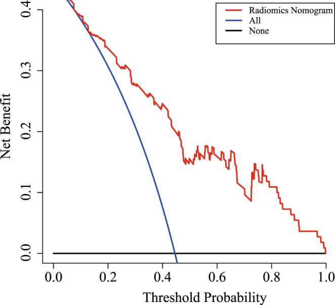
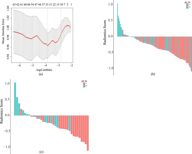
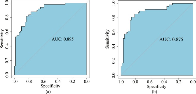
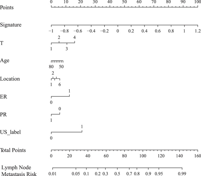
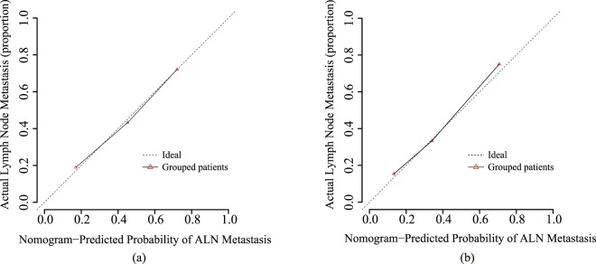
It is difficult to accurately assess axillary lymph nodes metastasis and the diagnosis of axillary lymph nodes in patients with breast cancer is invasive and has low-sensitivity preoperatively. This study aims to develop a mammography-based radiomics nomogram for the preoperative prediction of ALN metastasis in patients with breast cancer. This study enrolled 147 patients with clinicopathologically confirmed breast cancer and preoperative mammography. Features were extracted from each patient's mammography images. The least absolute shrinkage and selection operator regression method was used to select features and build a signature in the primary cohort. The performance of the signature was assessed using support vector machines. We developed a nomogram by incorporating the signature with the clinicopathologic risk factors. The nomogram performance was estimated by its calibration ability in the primary and validation cohorts. The signature was consisted of 10 selected ALN-status-related features. The AUC of the signature from the primary cohort was 0.895 (95% CI, 0.887-0.909) and 0.875 (95% CI, 0.698-0.891) for the validation cohort. The C-Index of the nomogram from the primary cohort was 0.779 (95% CI, 0.752-0.793) and 0.809 (95% CI, 0.794-0.833) for the validation cohort. Our nomogram is a reliable and non-invasive tool for preoperative prediction of ALN status and can be used to optimize current treatment strategy for breast cancer patients.

摘要

评估乳腺癌患者腋窝淋巴结转移和诊断腋窝淋巴结具有一定难度,且术前具有侵袭性和低敏感性。本研究旨在开发一种基于乳腺 X 线摄影术的放射组学列线图,用于预测乳腺癌患者腋窝淋巴结转移。本研究纳入了 147 例经临床病理证实的乳腺癌患者和术前乳腺 X 线摄影术。从每位患者的乳腺 X 线摄影图像中提取特征。采用最小绝对收缩和选择算子回归方法在主要队列中选择特征并构建特征签名。采用支持向量机评估特征签名的性能。我们通过将特征签名与临床病理危险因素相结合,开发了一个列线图。通过在主要和验证队列中评估其校准能力来估计列线图的性能。特征签名由 10 个选定的与腋窝淋巴结状态相关的特征组成。主要队列中特征签名的 AUC 为 0.895(95%CI,0.887-0.909),验证队列为 0.875(95%CI,0.698-0.891)。主要队列中列线图的 C-Index 为 0.779(95%CI,0.752-0.793),验证队列为 0.809(95%CI,0.794-0.833)。我们的列线图是一种用于预测腋窝淋巴结状态的可靠、非侵入性工具,可用于优化当前乳腺癌患者的治疗策略。

https://cdn.ncbi.nlm.nih.gov/pmc/blobs/01f3/6418289/65e1f85e37f9/41598_2019_40831_Fig6_HTML.jpg
https://cdn.ncbi.nlm.nih.gov/pmc/blobs/01f3/6418289/a55a1e26c082/41598_2019_40831_Fig1_HTML.jpg
https://cdn.ncbi.nlm.nih.gov/pmc/blobs/01f3/6418289/dd7aeeccd0f2/41598_2019_40831_Fig2_HTML.jpg
https://cdn.ncbi.nlm.nih.gov/pmc/blobs/01f3/6418289/ab983228b90c/41598_2019_40831_Fig3_HTML.jpg
https://cdn.ncbi.nlm.nih.gov/pmc/blobs/01f3/6418289/ffb7022f7850/41598_2019_40831_Fig4_HTML.jpg
https://cdn.ncbi.nlm.nih.gov/pmc/blobs/01f3/6418289/2cd8871c7ca0/41598_2019_40831_Fig5_HTML.jpg
https://cdn.ncbi.nlm.nih.gov/pmc/blobs/01f3/6418289/65e1f85e37f9/41598_2019_40831_Fig6_HTML.jpg

相似文献

1
Preoperative Prediction of Axillary Lymph Node Metastasis in Breast Cancer Using Mammography-Based Radiomics Method.基于乳腺 X 线摄影的放射组学方法预测乳腺癌腋窝淋巴结转移
Sci Rep. 2019 Mar 14;9(1):4429. doi: 10.1038/s41598-019-40831-z.
2
Mammography-based radiomics nomogram: a potential biomarker to predict axillary lymph node metastasis in breast cancer.基于乳腺 X 线摄影的放射组学列线图:一种预测乳腺癌腋窝淋巴结转移的潜在生物标志物。
Br J Radiol. 2020 Jul;93(1111):20191019. doi: 10.1259/bjr.20191019. Epub 2020 May 27.
3
Ultrasound-based radiomics nomogram: A potential biomarker to predict axillary lymph node metastasis in early-stage invasive breast cancer.基于超声的放射组学列线图:预测早期浸润性乳腺癌腋窝淋巴结转移的潜在生物标志物。
Eur J Radiol. 2019 Oct;119:108658. doi: 10.1016/j.ejrad.2019.108658. Epub 2019 Sep 7.
4
A two-center study of a combined nomogram based on mammography and MRI to predict ALN metastasis in breast cancer.基于乳腺 X 线摄影和 MRI 的联合列线图预测乳腺癌前哨淋巴结转移的两中心研究。
Magn Reson Imaging. 2024 Jul;110:128-137. doi: 10.1016/j.mri.2024.04.019. Epub 2024 Apr 15.
5
Non-invasive prediction model of axillary lymph node status in patients with early-stage breast cancer: a feasibility study based on dynamic contrast-enhanced-MRI radiomics.基于动态对比增强 MRI 放射组学的早期乳腺癌患者腋窝淋巴结状态的无创预测模型:一项可行性研究。
Br J Radiol. 2024 Feb 2;97(1154):439-450. doi: 10.1093/bjr/tqad034.
6
Magnetic resonance imaging radiomics predicts preoperative axillary lymph node metastasis to support surgical decisions and is associated with tumor microenvironment in invasive breast cancer: A machine learning, multicenter study.磁共振成像放射组学预测术前腋窝淋巴结转移以支持手术决策,并与浸润性乳腺癌的肿瘤微环境相关:一项机器学习、多中心研究。
EBioMedicine. 2021 Jul;69:103460. doi: 10.1016/j.ebiom.2021.103460. Epub 2021 Jul 4.
7
Multi-modality radiomics model predicts axillary lymph node metastasis of breast cancer using MRI and mammography.多模态放射组学模型利用 MRI 和乳腺 X 线摄影预测乳腺癌腋窝淋巴结转移。
Eur Radiol. 2024 Sep;34(9):6121-6131. doi: 10.1007/s00330-024-10638-2. Epub 2024 Feb 10.
8
Development and Validation of a Preoperative Magnetic Resonance Imaging Radiomics-Based Signature to Predict Axillary Lymph Node Metastasis and Disease-Free Survival in Patients With Early-Stage Breast Cancer.基于术前磁共振成像放射组学的signature 模型:预测早期乳腺癌患者腋窝淋巴结转移和无病生存的研究
JAMA Netw Open. 2020 Dec 1;3(12):e2028086. doi: 10.1001/jamanetworkopen.2020.28086.
9
Dynamic contrast-enhanced MRI radiomics nomogram for predicting axillary lymph node metastasis in breast cancer.动态对比增强 MRI 放射组学列线图预测乳腺癌腋窝淋巴结转移。
Cancer Imaging. 2022 Apr 4;22(1):17. doi: 10.1186/s40644-022-00450-w.
10
Radiomics model based on shear-wave elastography in the assessment of axillary lymph node status in early-stage breast cancer.基于剪切波弹性成像的放射组学模型在早期乳腺癌腋窝淋巴结状态评估中的应用。
Eur Radiol. 2022 Apr;32(4):2313-2325. doi: 10.1007/s00330-021-08330-w. Epub 2021 Oct 20.

引用本文的文献

1
Contrast-enhanced ultrasound radiomics model for predicting axillary lymph node metastasis and prognosis in breast cancer: a multicenter study.用于预测乳腺癌腋窝淋巴结转移及预后的超声造影放射组学模型:一项多中心研究
BMC Cancer. 2025 Aug 14;25(1):1315. doi: 10.1186/s12885-025-14632-9.
2
Deep learning on routine full-breast mammograms enhances lymph node metastasis prediction in early breast cancer.基于常规全乳钼靶X线摄影的深度学习可提高早期乳腺癌淋巴结转移的预测能力。
NPJ Digit Med. 2025 Jul 10;8(1):425. doi: 10.1038/s41746-025-01831-8.
3
Development and validation of a multivariable risk model based on clinicopathological characteristics, mammography, and MRI imaging features for predicting axillary lymph node metastasis in patients with upgraded ductal carcinoma .

本文引用的文献

1
Preoperative assessment of axillary lymph node status in breast cancer patients by ultrasonography combined with mammography: A STROBE compliant article.超声检查联合乳腺钼靶对乳腺癌患者腋窝淋巴结状态的术前评估:一篇遵循STROBE规范的文章
Medicine (Baltimore). 2018 Jul;97(30):e11441. doi: 10.1097/MD.0000000000011441.
2
Preoperative prediction nomogram based on primary tumor miRNAs signature and clinical-related features for axillary lymph node metastasis in early-stage invasive breast cancer.基于原发性肿瘤 miRNA 特征和临床相关特征的早期浸润性乳腺癌腋窝淋巴结转移的术前预测列线图。
Int J Cancer. 2018 May 1;142(9):1901-1910. doi: 10.1002/ijc.31208. Epub 2018 Jan 12.
3
基于临床病理特征、乳腺钼靶和MRI成像特征的多变量风险模型的开发与验证,用于预测升级型导管癌患者的腋窝淋巴结转移
Gland Surg. 2025 Apr 30;14(4):738-753. doi: 10.21037/gs-2025-89. Epub 2025 Apr 25.
4
Clinical-radiomics model for predicting internal mammary lymph node metastasis in operable breast cancer patients.用于预测可手术乳腺癌患者内乳淋巴结转移的临床影像组学模型
Front Oncol. 2025 Apr 3;15:1477866. doi: 10.3389/fonc.2025.1477866. eCollection 2025.
5
Enhancing Specificity in Predicting Axillary Lymph Node Metastasis in Breast Cancer through an Interpretable Machine Learning Model with CEM and Ultrasound Integration.通过具有CEM和超声整合的可解释机器学习模型提高乳腺癌腋窝淋巴结转移预测的特异性
Technol Cancer Res Treat. 2025 Jan-Dec;24:15330338251334735. doi: 10.1177/15330338251334735. Epub 2025 Apr 17.
6
Prediction of axillary lymph node metastasis in breast cancer patients based on ultrasonograhic-clinicopathologic features.基于超声-临床病理特征预测乳腺癌患者腋窝淋巴结转移
Pak J Med Sci. 2025 Jan;41(1):96-100. doi: 10.12669/pjms.41.1.10384.
7
Identification of sentinel lymph node macrometastasis in breast cancer by deep learning based on clinicopathological characteristics.基于临床病理特征的深度学习在乳腺癌前哨淋巴结宏转移中的鉴定。
Sci Rep. 2024 Nov 6;14(1):26970. doi: 10.1038/s41598-024-78040-y.
8
Preoperative prediction of nodal status using clinical data and artificial intelligence derived mammogram features enabling abstention of sentinel lymph node biopsy in breast cancer.利用临床数据和人工智能提取的乳房X光检查特征进行术前淋巴结状态预测,可避免乳腺癌患者进行前哨淋巴结活检。
Front Oncol. 2024 Jul 10;14:1394448. doi: 10.3389/fonc.2024.1394448. eCollection 2024.
9
Ultrasound-based radiomics nomogram for predicting axillary lymph node metastasis in invasive breast cancer.基于超声的影像组学列线图预测浸润性乳腺癌腋窝淋巴结转移
Am J Transl Res. 2024 Jun 15;16(6):2398-2410. doi: 10.62347/KEPZ9726. eCollection 2024.
10
Assessment of the axilla in women with early-stage breast cancer undergoing primary surgery: a review.早期乳腺癌行原发性手术的女性腋窝评估:综述。
World J Surg Oncol. 2024 May 9;22(1):127. doi: 10.1186/s12957-024-03394-6.
Biomarkers and Imaging of Breast Cancer.
乳腺癌的生物标志物和影像学诊断
AJR Am J Roentgenol. 2018 Feb;210(2):271-278. doi: 10.2214/AJR.17.18708. Epub 2017 Nov 22.
4
Sentinel Lymph Node Biopsy in Breast Cancer: A Clinical Review and Update.乳腺癌前哨淋巴结活检:临床综述与更新
J Breast Cancer. 2017 Sep;20(3):217-227. doi: 10.4048/jbc.2017.20.3.217. Epub 2017 Sep 22.
5
Radiomics Analysis for Evaluation of Pathological Complete Response to Neoadjuvant Chemoradiotherapy in Locally Advanced Rectal Cancer.放射组学分析评估局部晚期直肠癌新辅助放化疗的病理完全缓解。
Clin Cancer Res. 2017 Dec 1;23(23):7253-7262. doi: 10.1158/1078-0432.CCR-17-1038. Epub 2017 Sep 22.
6
A Radiomics Nomogram for the Preoperative Prediction of Lymph Node Metastasis in Bladder Cancer.膀胱癌淋巴结转移术前预测的放射组学列线图
Clin Cancer Res. 2017 Nov 15;23(22):6904-6911. doi: 10.1158/1078-0432.CCR-17-1510. Epub 2017 Sep 5.
7
Preoperative prediction of sentinel lymph node metastasis in breast cancer based on radiomics of T2-weighted fat-suppression and diffusion-weighted MRI.基于 T2 压脂和弥散加权 MRI 放射组学的乳腺癌前哨淋巴结转移术前预测。
Eur Radiol. 2018 Feb;28(2):582-591. doi: 10.1007/s00330-017-5005-7. Epub 2017 Aug 21.
8
[F]FDG PET/CT features for the molecular characterization of primary breast tumors.[F]FDG PET/CT 特征用于原发性乳腺癌的分子特征分析。
Eur J Nucl Med Mol Imaging. 2017 Nov;44(12):1945-1954. doi: 10.1007/s00259-017-3770-9. Epub 2017 Jul 15.
9
Prediction of nonsentinel lymph node metastasis in breast cancer patients with one or two positive sentinel lymph nodes.预测一个或两个前哨淋巴结阳性的乳腺癌患者的非前哨淋巴结转移。
Asian J Surg. 2018 Jan;41(1):12-19. doi: 10.1016/j.asjsur.2016.06.001. Epub 2016 Aug 31.
10
Development and Validation of a Radiomics Nomogram for Preoperative Prediction of Lymph Node Metastasis in Colorectal Cancer.基于影像组学的直肠癌淋巴结转移术前预测列线图模型的建立与验证。
J Clin Oncol. 2016 Jun 20;34(18):2157-64. doi: 10.1200/JCO.2015.65.9128. Epub 2016 May 2.