Chen Jun-Hui, Wu Ting, Xia Wen-Yuan, Shi Zhong-Hua, Zhang Chun-Lei, Chen Lei, Chen Qian-Xue, Wang Yu-Hai
Department of Neurosurgery, Renmin Hospital of Wuhan University, Wuhan, Hubei Province; Department of Neurosurgery, Wuxi Clinical College of Anhui Medical University, 904 Hospital of Joint Logistic Support Force of PLA, Wuxi, Jiangsu Province, China.
Department of Cardiology, Wuxi Clinical College of Anhui Medical University, 904 Hospital of Joint Logistic Support Force of PLA, Wuxi, Jiangsu Province, China.
Neural Regen Res. 2020 Oct;15(10):1947-1954. doi: 10.4103/1673-5374.280326.
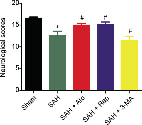
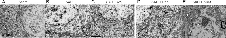
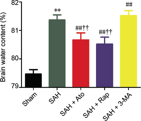
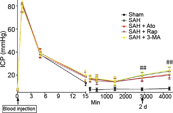
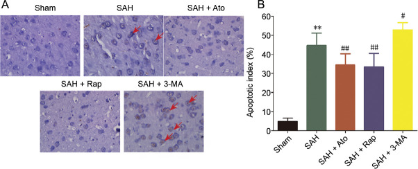
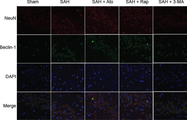
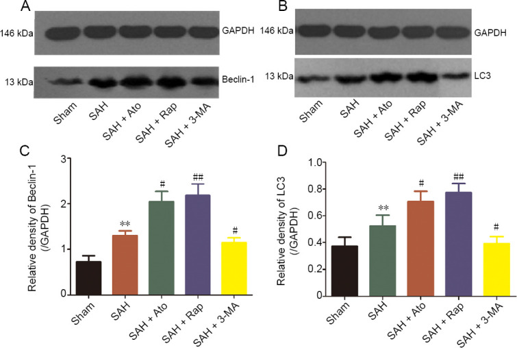
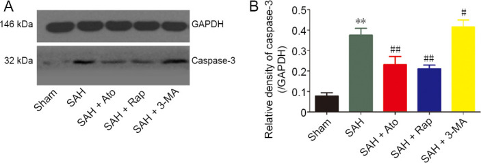
Atorvastatin has been shown to reduce early brain edema and neuronal death after subarachnoid hemorrhage, but its mechanism is not clear. In this study, rat models of subarachnoid hemorrhage were established by autologous blood injection in the cisterna magna. Rat models were intragastrically administered 20 mg/kg atorvastatin 24 hours before subarachnoid hemorrhage, 12 and 36 hours after subarachnoid hemorrhage. Compared with the controls, atorvastatin treatment demonstrated that at 72 hours after subarachnoid hemorrhage, neurological function had clearly improved; brain edema was remarkably relieved; cell apoptosis was markedly reduced in the cerebral cortex of rats; the number of autophagy-related protein Beclin-1-positive cells and the expression levels of Beclin-1 and LC3 were increased compared with subarachnoid hemorrhage only. The ultrastructural damage of neurons in the temporal lobe was also noticeably alleviated. The similarities between the effects of atorvastatin and rapamycin were seen in all the measured outcomes of subarachnoid hemorrhage. However, these were contrary to the results of 3-methyladenine injection, which inhibits the signaling pathway of autophagy. These findings indicate that atorvastatin plays an early neuroprotective role in subarachnoid hemorrhage by activating autophagy. The experimental protocol was approved by the Animal Ethics Committee of Anhui Medical University, China (904 Hospital of Joint Logistic Support Force of PLA; approval No. YXLL-2017-09) on February 22, 2017.
阿托伐他汀已被证明可减轻蛛网膜下腔出血后的早期脑水肿和神经元死亡,但其机制尚不清楚。在本研究中,通过在大鼠小脑延髓池注射自体血建立蛛网膜下腔出血大鼠模型。在蛛网膜下腔出血前24小时、蛛网膜下腔出血后12小时和36小时,对大鼠模型进行灌胃给予20mg/kg阿托伐他汀。与对照组相比,阿托伐他汀治疗显示,在蛛网膜下腔出血72小时后,神经功能明显改善;脑水肿明显减轻;大鼠大脑皮质细胞凋亡明显减少;与单纯蛛网膜下腔出血相比,自噬相关蛋白Beclin-1阳性细胞数量以及Beclin-1和LC3的表达水平增加。颞叶神经元的超微结构损伤也明显减轻。在蛛网膜下腔出血的所有测量结果中,阿托伐他汀和雷帕霉素的作用相似。然而,这些结果与抑制自噬信号通路的3-甲基腺嘌呤注射结果相反。这些发现表明,阿托伐他汀通过激活自噬在蛛网膜下腔出血中发挥早期神经保护作用。该实验方案于2017年2月22日获得中国安徽医科大学动物伦理委员会(中国人民解放军联勤保障部队第904医院;批准号YXLL-2017-09)的批准。