Di Jia, Liu Yuqiu, Wang Dongyan, Yang Min
Nephrology, Department of Nephrology, Changzhou First People's Hospital, Changzhou, China.
Institute of Nephrology, Institute of Nephrology, Zhong Da Hospital, Southeast University, School of Medicine, Nanjing, Jiangsu, China.
Case Rep Med. 2020 Mar 17;2020:9526836. doi: 10.1155/2020/9526836. eCollection 2020.
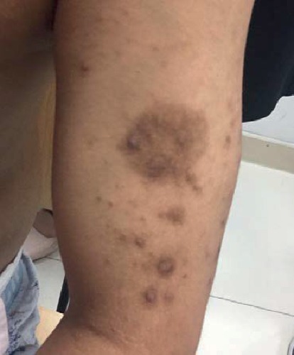
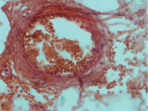
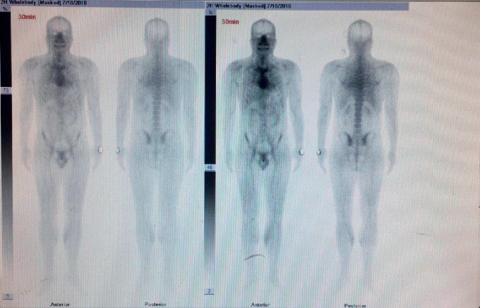
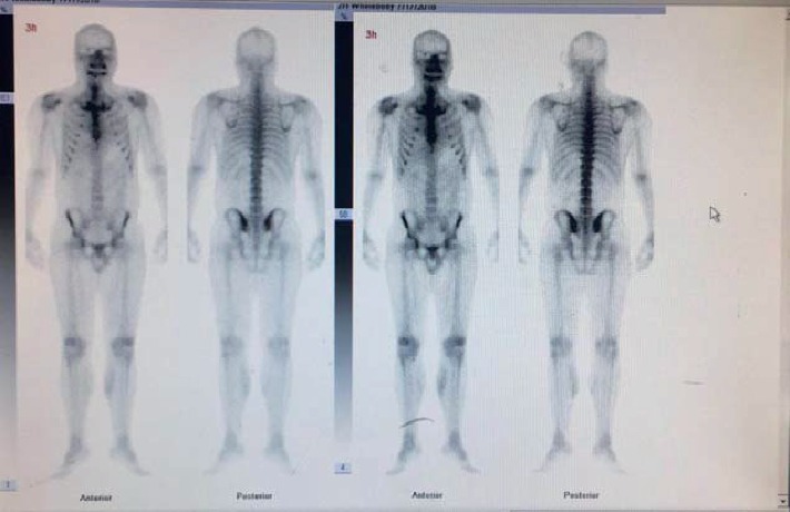
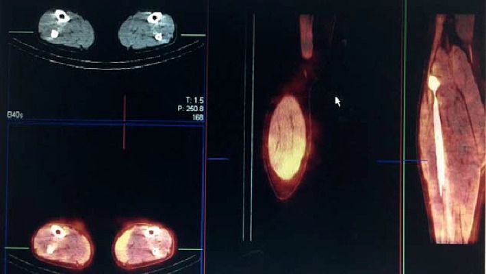
Calcium uremic aortic disease (calciphylaxis) has long been considered as a rare, life-threatening small vessel disease. The diagnosis of calciphylaxis depends mainly on clinical symptoms and high risk factors, and skin biopsy can be used to confirm the diagnosis. However, noninvasive testing methods are still the focus of exploration currently. There is increasing evidence that bone scintigraphy is helpful in the diagnosis of calciphylaxis, especially for assessing the involvement of muscles and internal organs. Here, we describe a pathology-proven case of calciphylaxis case and the corresponding imaging findings on Tc-99 m MDP bone scan imaging.
钙性尿毒症性主动脉疾病(钙化防御)长期以来一直被认为是一种罕见的、危及生命的小血管疾病。钙化防御的诊断主要依赖于临床症状和高危因素,皮肤活检可用于确诊。然而,无创检测方法仍是目前探索的重点。越来越多的证据表明,骨闪烁显像有助于钙化防御的诊断,尤其是在评估肌肉和内脏受累情况方面。在此,我们描述一例经病理证实的钙化防御病例以及相应的锝-99m亚甲基二膦酸盐(Tc-99 m MDP)骨扫描成像表现。