• 文献检索
  • 文档翻译
  • 深度研究
  • 学术资讯
  • Suppr Zotero 插件Zotero 插件
  • 邀请有礼
  • 套餐&价格
  • 历史记录
应用&插件
Suppr Zotero 插件Zotero 插件浏览器插件Mac 客户端Windows 客户端微信小程序
定价
高级版会员购买积分包购买API积分包
服务
文献检索文档翻译深度研究API 文档MCP 服务
关于我们
关于 Suppr公司介绍联系我们用户协议隐私条款
关注我们

Suppr 超能文献

核心技术专利:CN118964589B侵权必究
粤ICP备2023148730 号-1Suppr @ 2026

文献检索

告别复杂PubMed语法,用中文像聊天一样搜索,搜遍4000万医学文献。AI智能推荐,让科研检索更轻松。

立即免费搜索

文件翻译

保留排版,准确专业,支持PDF/Word/PPT等文件格式,支持 12+语言互译。

免费翻译文档

深度研究

AI帮你快速写综述,25分钟生成高质量综述,智能提取关键信息,辅助科研写作。

立即免费体验

灌注计算机断层扫描成像在腮腺良性与恶性病变鉴别中的应用

Perfusion Computed Tomography Scan Imaging in Differentiation of Benign from Malignant Parotid Lesions.

作者信息

Niazi Masume, Mohammadzadeh Maryam, Aghazadeh Kayvan, Sharifian Hashem, Karimi Ebrahim, Shakiba Madjid, Baniasadi Maria, Rahmaty Benyamin, Adel Suzan, Moharreri Melorina

机构信息

Department of Otorhinolaryngology, Otorhinolaryngology Research Center, Tehran University of Medical Sciences, Tehran, Iran.

Division of Neuroradiology, Department of Radiology, Amir Aalam Hospital, Tehran University of Medical Sciences, Tehran, Iran.

出版信息

Int Arch Otorhinolaryngol. 2020 Apr;24(2):e160-e169. doi: 10.1055/s-0039-1697005. Epub 2019 Nov 4.

DOI:10.1055/s-0039-1697005
PMID:32256836
原文链接:https://pmc.ncbi.nlm.nih.gov/articles/PMC6828566/
Abstract

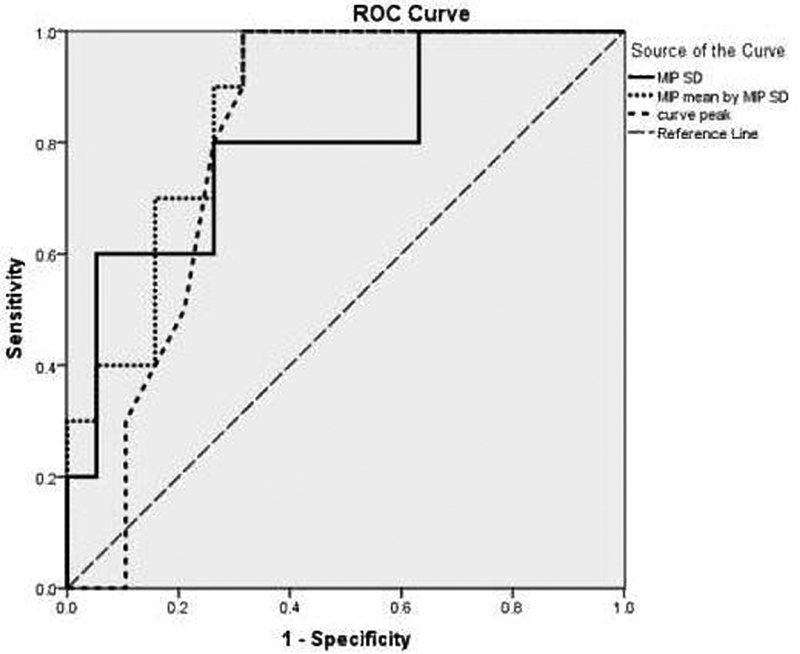
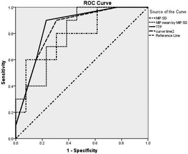
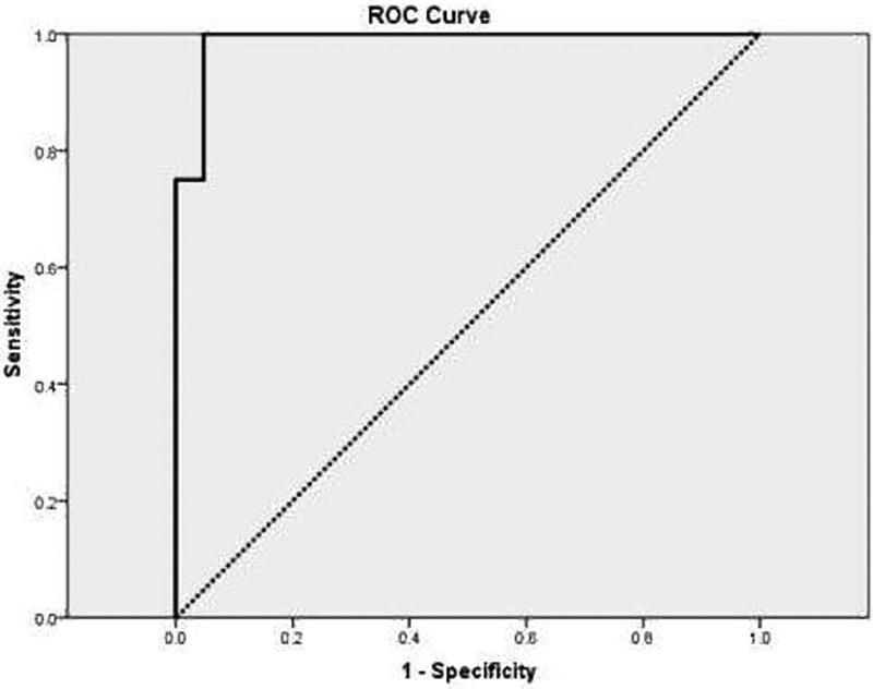
The most common site of salivary gland tumors is the parotid gland. Computed tomography (CT), magnetic resonance imaging (MRI), and sonography are imaging modalities to differentiate benign from malignant parotid tumors.  The aim of this study is the evaluation of the diagnostic value of perfusion CT for differentiating histological categorization of benign and malignant parotid tumors.  A total of 29 patients with parotid neoplasms were enrolled in this study. Mean age and all CT perfusion variables (gradient and permeability, blood flow [BF], blood volume [BV], mean transit time [MTT], permeability surface [PS], maximum intensity projection [MIP], time-density curve [TDC], and time to peak [TTP]) were compared among three groups (malignant tumors [MTs], Warthin's tumor [WT] and pleomorphic adenomas [PA]).  The mean age of the patients was 55.9 ± 14.1 (26-77), and 15 of them were male (51.7%). Eleven lesions were PAs [37.9%], 8 lesions were WTs (27.6%0 and 10 lesions (34.5%) were MTs (6 acinic cell carcinomas [ACCs], 3 adenocystic carcinomas [AdCCs], and 1 mucoepidermoid carcinoma [MEC]). The mean age of the patients with WTs was 62 ± 7.5 years; 52 ± 14.2 for patients with Pas, and 55.2 ± 17.2 for those with MTs (  = 0.32). The mean MIP was 122.7 ± 12.2 in WT, while it was 80.5 ± 19.5 in PA, and 76.2 ± 27.1 in MTs (  < 0.001); The mean MIP for WT was higher than for PAs and MTs; the values of MTs and PAs were not statistically different. The average of BF, BV, and curve peak were higher in WTs in comparison with the other two groups, and curve time 2 and TTP were higher in PAs in comparison with MTs.  Based on this study, perfusion CT of the parotid gland and its parameters can distinguish between benign and malignant parotid masses.

摘要

涎腺肿瘤最常见的发生部位是腮腺。计算机断层扫描(CT)、磁共振成像(MRI)和超声检查是区分腮腺良恶性肿瘤的影像学检查方法。 本研究的目的是评估CT灌注成像在鉴别腮腺良恶性肿瘤组织学分类中的诊断价值。 本研究共纳入29例腮腺肿瘤患者。比较了三组(恶性肿瘤[MTs]、沃辛瘤[WT]和多形性腺瘤[PA])患者的平均年龄以及所有CT灌注参数(梯度和通透性、血流量[BF]、血容量[BV]、平均通过时间[MTT]、通透表面积[PS]、最大密度投影[MIP]、时间-密度曲线[TDC]和达峰时间[TTP])。 患者的平均年龄为55.9±14.1岁(26 - 77岁),其中15例为男性(51.7%)。11个病灶为多形性腺瘤[37.9%],8个病灶为沃辛瘤(27.6%),10个病灶(34.5%)为恶性肿瘤(6例腺泡细胞癌[ACCs]、3例腺样囊性癌[AdCCs]和1例黏液表皮样癌[MEC])。沃辛瘤患者的平均年龄为62±7.5岁;多形性腺瘤患者为52±14.2岁,恶性肿瘤患者为55.2±17.2岁(P = 0.32)。沃辛瘤的平均最大密度投影(MIP)为122.7±12.2,多形性腺瘤为80.5±19.5,恶性肿瘤为76.2±27.1(P < 0.001);沃辛瘤的平均MIP高于多形性腺瘤和恶性肿瘤;恶性肿瘤和多形性腺瘤的值无统计学差异。与其他两组相比,沃辛瘤的血流量、血容量和曲线峰值的平均值更高,与恶性肿瘤相比,多形性腺瘤的曲线时间2和达峰时间更高。 基于本研究,腮腺CT灌注成像及其参数可区分腮腺肿块的良恶性。

https://cdn.ncbi.nlm.nih.gov/pmc/blobs/c4dc/6828566/f04868f04eab/10-1055-s-0039-1697005-i0936or-3.jpg
https://cdn.ncbi.nlm.nih.gov/pmc/blobs/c4dc/6828566/0b65ea0ce058/10-1055-s-0039-1697005-i0936or-1.jpg
https://cdn.ncbi.nlm.nih.gov/pmc/blobs/c4dc/6828566/7426866105ea/10-1055-s-0039-1697005-i0936or-2.jpg
https://cdn.ncbi.nlm.nih.gov/pmc/blobs/c4dc/6828566/f04868f04eab/10-1055-s-0039-1697005-i0936or-3.jpg

相似文献

1
Perfusion Computed Tomography Scan Imaging in Differentiation of Benign from Malignant Parotid Lesions.灌注计算机断层扫描成像在腮腺良性与恶性病变鉴别中的应用
Int Arch Otorhinolaryngol. 2020 Apr;24(2):e160-e169. doi: 10.1055/s-0039-1697005. Epub 2019 Nov 4.
2
The Clinical Characteristics and CT Findings of Parotid and Submandibular Gland Tumours.腮腺及颌下腺肿瘤的临床特征与CT表现
J Oncol. 2021 Jul 3;2021:8874100. doi: 10.1155/2021/8874100. eCollection 2021.
3
Tumor blood flow and apparent diffusion coefficient histogram analysis for differentiating malignant salivary tumors from pleomorphic adenomas and Warthin's tumors.肿瘤血流和表观扩散系数直方图分析用于鉴别恶性唾液腺肿瘤、多形性腺瘤和沃辛瘤。
Sci Rep. 2022 Apr 8;12(1):5947. doi: 10.1038/s41598-022-09968-2.
4
Influence of post-label delay time on the performance of 3D pseudo-continuous arterial spin labeling magnetic resonance imaging in the characterization of parotid gland tumors.基于 3D 伪连续动脉自旋标记磁共振成像后标记延迟时间对腮腺肿瘤特征分析的影响。
Eur Radiol. 2022 Feb;32(2):1087-1094. doi: 10.1007/s00330-021-08220-1. Epub 2021 Aug 4.
5
Intravoxel Incoherent Motion Magnetic Resonance Imaging for Assessing Parotid Gland Tumors: Correlation and Comparison with Arterial Spin Labeling Imaging.体素内不相干运动磁共振成像评估腮腺肿瘤:与动脉自旋标记成像的相关性和比较。
Korean J Radiol. 2021 Feb;22(2):243-252. doi: 10.3348/kjr.2020.0290. Epub 2020 Sep 10.
6
Differentiation of benign and malignant parotid tumors using deconvolution-based perfusion CT imaging: feasibility of the method and initial results.基于去卷积的灌注CT成像鉴别腮腺良恶性肿瘤:该方法的可行性及初步结果
Eur J Radiol. 2007 Nov;64(2):258-65. doi: 10.1016/j.ejrad.2007.02.032. Epub 2007 Mar 30.
7
Diagnostic value of maximum signal intensity on T1-weighted MRI images for differentiating parotid gland tumours along with pathological correlation.T1 加权 MRI 图像上最大信号强度对鉴别腮腺肿瘤的诊断价值及病理相关性分析。
Clin Radiol. 2021 Jun;76(6):472.e19-472.e25. doi: 10.1016/j.crad.2021.02.011. Epub 2021 Mar 14.
8
Conventional, diffusion, and dynamic contrast-enhanced MRI findings for differentiating metaplastic Warthin's tumor of the parotid gland.常规、扩散和动态对比增强 MRI 对腮腺化生性 Warthin 肿瘤的鉴别诊断。
Sci Prog. 2021 Apr-Jun;104(2):368504211018583. doi: 10.1177/00368504211018583.
9
Quantitative dynamic contrast-enhanced MRI and readout segmentation of long variable echo-trains diffusion-weighted imaging in differentiating parotid gland tumors.定量动态对比增强磁共振成像和读出分段长变量回波扩散加权成像在鉴别腮腺肿瘤中的应用。
Neuroradiology. 2021 Oct;63(10):1709-1719. doi: 10.1007/s00234-021-02758-z. Epub 2021 Jul 9.
10
Contrast-Enhanced Ultrasound in the Differentiation between the Most Common Benign Parotid Gland Tumors: A Systematic Review and Meta-Analysis.超声造影在常见腮腺良性肿瘤鉴别诊断中的应用:一项系统评价与Meta分析
J Clin Med. 2022 Dec 12;11(24):7360. doi: 10.3390/jcm11247360.

引用本文的文献

1
Diffusion-derived vessel density (DDVD) for penumbra delineation in brain acute ischemic stroke: initial proof-of-concept results using single NEX DWI.用于脑急性缺血性卒中半暗带勾画的扩散衍生血管密度(DDVD):使用单次激励扩散加权成像的初步概念验证结果
Quant Imaging Med Surg. 2024 Dec 5;14(12):9533-9542. doi: 10.21037/qims-24-2139. Epub 2024 Oct 21.
2
Survival in Patients with Primary Parotid Gland Carcinoma after Surgery-Results of a Single-Centre Study.原发腮腺腺癌患者手术后的生存情况——单中心研究结果。
Curr Oncol. 2023 Feb 23;30(3):2702-2714. doi: 10.3390/curroncol30030204.

本文引用的文献

1
State-of-the-Art Imaging of Salivary Gland Tumors.唾液腺肿瘤的最新成像技术
Neuroimaging Clin N Am. 2018 May;28(2):303-317. doi: 10.1016/j.nic.2018.01.009.
2
Imaging of Inflammatory Disorders of Salivary Glands.唾液腺炎症性疾病的影像学检查
Neuroimaging Clin N Am. 2018 May;28(2):255-272. doi: 10.1016/j.nic.2018.01.006.
3
Salivary gland pleomorphic adenoma in the Netherlands: A nationwide observational study of primary tumor incidence, malignant transformation, recurrence, and risk factors for recurrence.
荷兰唾液腺多形性腺瘤:一项关于原发性肿瘤发病率、恶变、复发及复发危险因素的全国性观察性研究。
Oral Oncol. 2017 Mar;66:93-99. doi: 10.1016/j.oraloncology.2017.01.004. Epub 2017 Jan 23.
4
Role of perfusion CT in the evaluation of functional primary tumour response after radiochemotherapy in head and neck cancer: preliminary findings.灌注CT在头颈部癌放化疗后功能性原发肿瘤反应评估中的作用:初步研究结果
Br J Radiol. 2016 Sep;89(1065):20151070. doi: 10.1259/bjr.20151070. Epub 2016 Jul 5.
5
Differential diagnostic value of computed tomography perfusion combined with vascular endothelial growth factor expression in head and neck lesions.计算机断层扫描灌注成像联合血管内皮生长因子表达在头颈部病变中的鉴别诊断价值
Oncol Lett. 2016 May;11(5):3342-3348. doi: 10.3892/ol.2016.4413. Epub 2016 Apr 5.
6
Effect of increasing the sampling interval to 2 seconds on the radiation dose and accuracy of CT perfusion of the head and neck.将采样间隔增加至2秒对头部和颈部CT灌注的辐射剂量及准确性的影响。
J Comput Assist Tomogr. 2014 May-Jun;38(3):469-73. doi: 10.1097/RCT.0000000000000066.
7
Perfusion CT of head and neck cancer.头颈部癌症的灌注 CT。
Eur J Radiol. 2014 Mar;83(3):537-44. doi: 10.1016/j.ejrad.2013.12.008. Epub 2013 Dec 16.
8
Diagnostic value of CT perfusion imaging for parotid neoplasms.CT 灌注成像对腮腺肿瘤的诊断价值。
Dentomaxillofac Radiol. 2014;43(1):20130237. doi: 10.1259/dmfr.20130237. Epub 2013 Nov 1.
9
Evaluation of cervical lymph nodes with CT perfusion in patients with hypopharyngeal and laryngeal squamous cell cancer.下咽及喉鳞状细胞癌患者颈部淋巴结的CT灌注评估
Pol J Radiol. 2011 Jan;76(1):7-13.
10
Head and neck cancer: value of perfusion CT in depicting primary tumor spread.头颈部癌:灌注 CT 在描绘原发性肿瘤扩散方面的价值。
Med Sci Monit. 2012 Feb;18(2):CR112-118. doi: 10.12659/msm.882466.