Robles-Medranda Carlos, Oleas Roberto, Valero Manuel, Puga-Tejada Miguel, Soria-Alcívar Miguel, Ospina Jesenia, Alvarado-Escobar Haydee, Muñoz-Jurado Guillermo, Baquerizo-Burgos Jorge, Pitanga-Lukashok Hannah
Gastroenterology and Endoscopy Division, Instituto Ecuatoriano de Enfermedades Digestivas (IECED), Guayaquil, Ecuador.
Endosc Int Open. 2020 Apr;8(4):E550-E557. doi: 10.1055/a-1119-6327. Epub 2020 Mar 23.
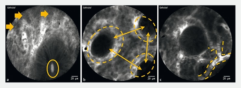
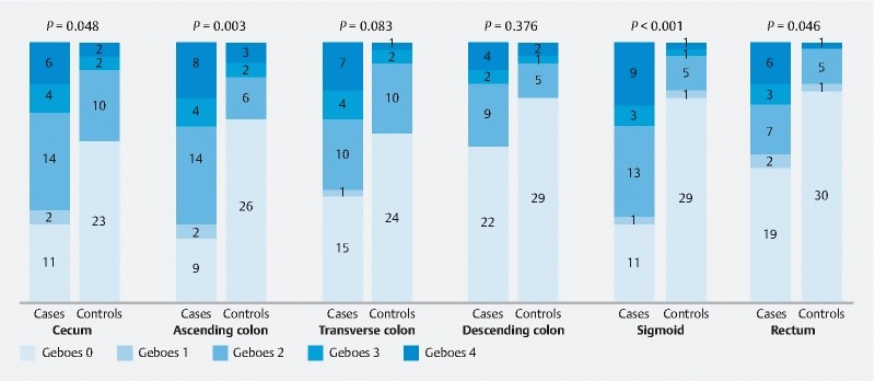
Irritable bowel syndrome (IBS) is considered to be a functional disease, but recent data indicate measurable organic alterations. We aimed to determine the presence of colorectal mucosa microinflammation in vivo via probe-confocal laser endomicroscopy (pCLE) and histological evaluation in IBS patients. This was a prospective, controlled, nonrandomized single-blind diagnostic trial performed in a tertiary institution. pCLE images and targeted biopsy of each colon segment obtained during colonoscopies of IBS patients and controls were analyzed for inflammatory changes. Biopsies were classified using the Geboes scale, and the odds ratio and overall diagnostic accuracy were calculated. During the 15-month study period, 37 patients were allocated to each group. The mean age was 53.1 ± 14.3 years; 64.9 % were female. Signs of colonic mucosa inflammation were evident on 65.8 % of pCLE images from IBS patients compared to 23.4 % of images from controls (OR 6.28; 4.14-9.52; < 0.001). In total, 20/37 patients had microinflammation via pCLE in ≥ 3 colon segments in the IBS group, compared to 1/37 in the control group. A Geboes score > 0 was attributed to 60.8 % of biopsies from patients in the IBS group compared to 27.5 % of biopsies from the control group. The sensitivity, specificity, positive and negative predictive values, observed and interrater agreement of pCLE-detected inflammatory changes in IBS using histology as gold standard were 76 %, 91 %, 76 %, 91 %, 86.5 %, and 66.8 %, respectively. Patients with IBS have a six-fold higher prevalence of colorectal mucosa microinflammation than healthy controls. pCLE might be a reliable method to detect colorectal mucosa microinflammation in IBS patients.
肠易激综合征(IBS)被认为是一种功能性疾病,但近期数据表明存在可测量的器质性改变。我们旨在通过探头共聚焦激光内镜检查(pCLE)和组织学评估来确定IBS患者体内结肠黏膜微炎症的存在情况。 这是一项在三级医疗机构进行的前瞻性、对照、非随机单盲诊断试验。对IBS患者和对照组结肠镜检查期间获得的每个结肠段的pCLE图像和靶向活检进行炎症变化分析。活检采用格博斯量表进行分类,并计算比值比和总体诊断准确性。 在15个月的研究期间,每组分配37例患者。平均年龄为53.1±14.3岁;64.9%为女性。IBS患者65.8%的pCLE图像显示结肠黏膜炎症迹象,而对照组图像为23.4%(比值比6.28;4.14 - 9.52;<0.001)。IBS组共有20/37例患者通过pCLE在≥3个结肠段存在微炎症,而对照组为1/37例。IBS组患者活检中有60.8%的格博斯评分>0,而对照组活检为27.5%。以组织学为金标准,pCLE检测IBS炎症变化的敏感性、特异性、阳性和阴性预测值、观察一致性和观察者间一致性分别为76%、91%、76%、91%、86.5%和66.8%。 IBS患者结肠黏膜微炎症的患病率比健康对照组高六倍。pCLE可能是检测IBS患者结肠黏膜微炎症的可靠方法。