Department of Chemistry, University of Texas, Austin, Texas 78712, United States.
Division of Chemical Biology and Medicinal Chemistry, University of Texas, Austin, Texas 78712, United States.
J Am Soc Mass Spectrom. 2020 May 6;31(5):1140-1150. doi: 10.1021/jasms.0c00066. Epub 2020 Apr 21.
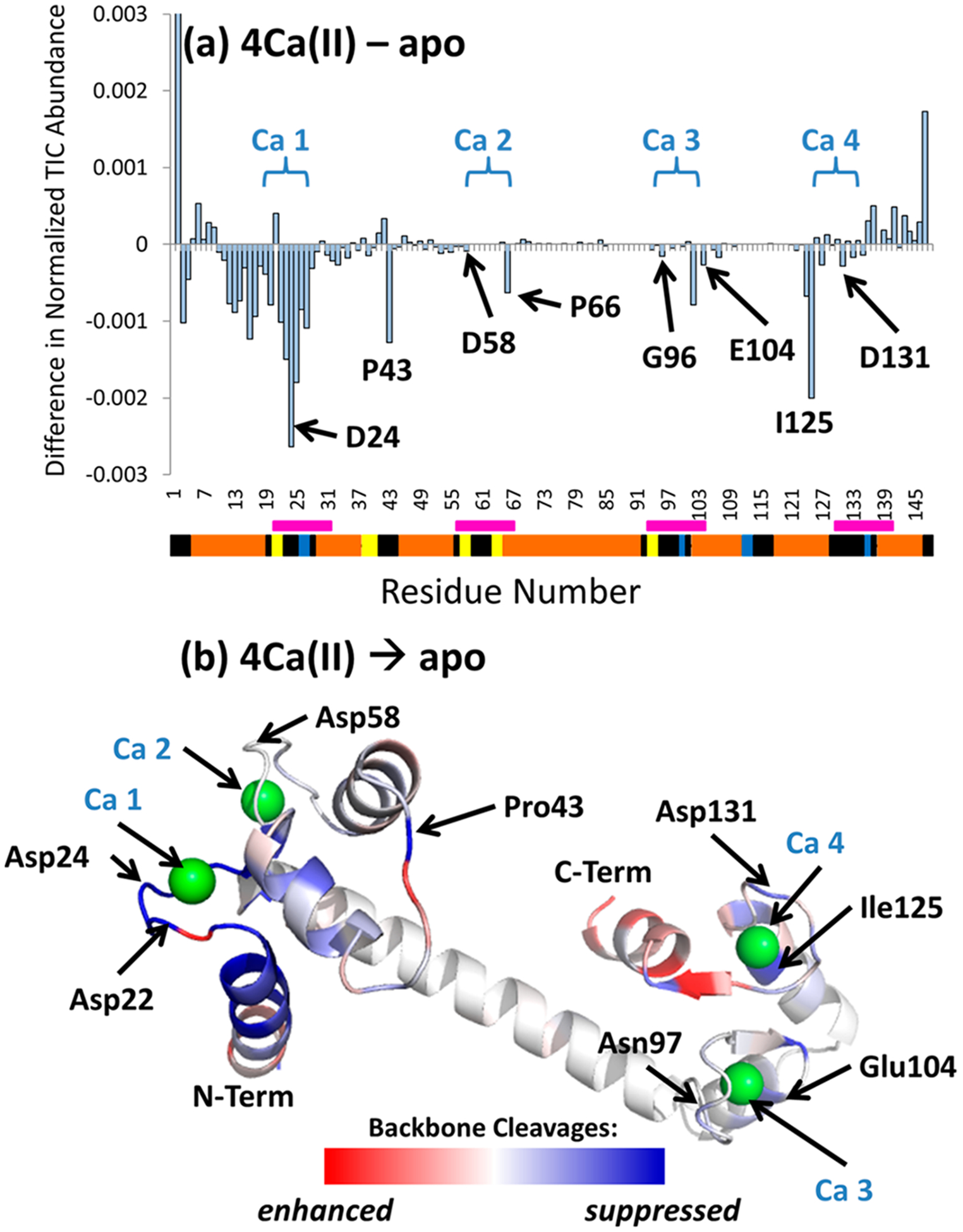
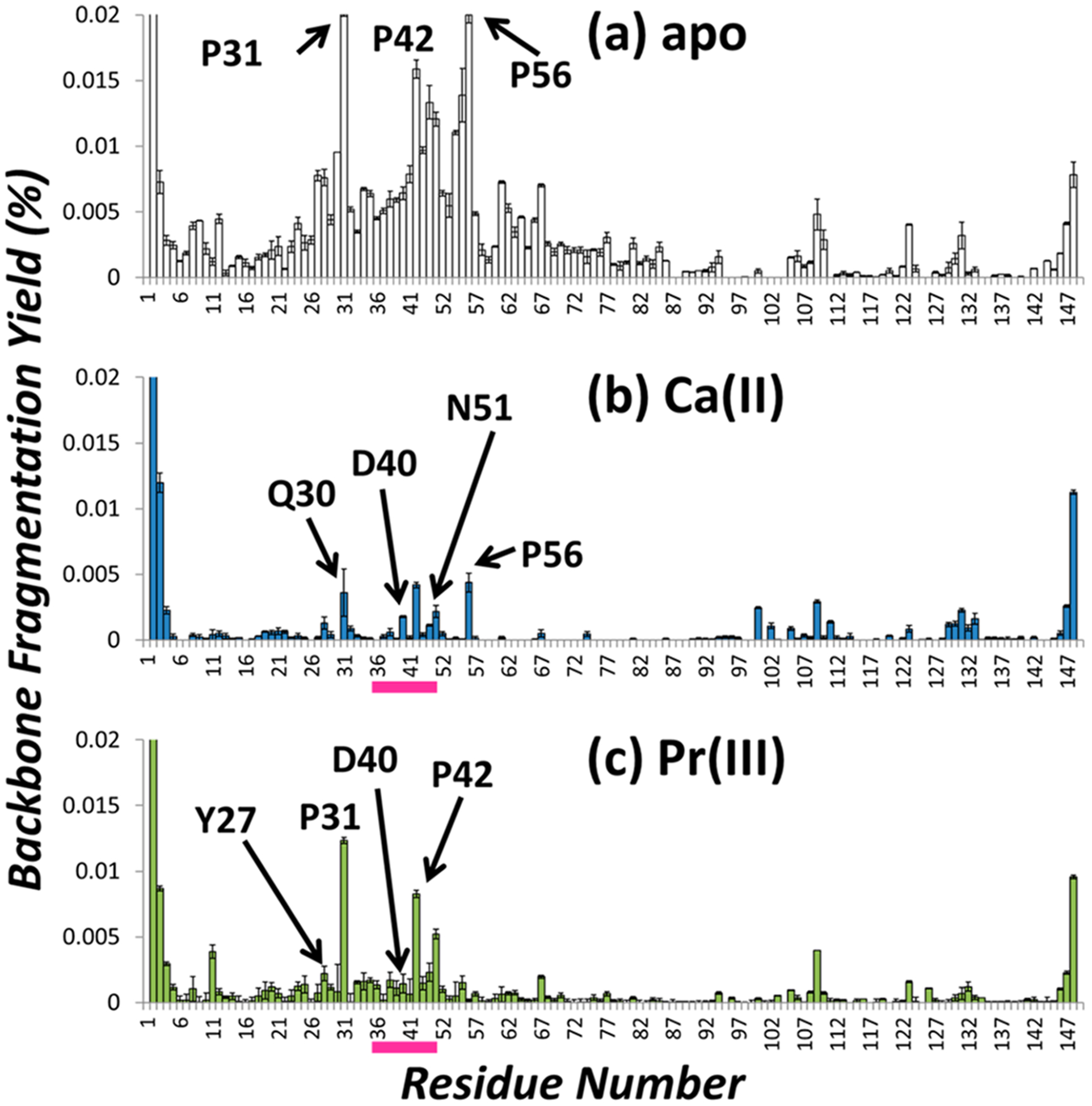
Ultraviolet photodissociation (UVPD) has emerged as a promising tool to characterize proteins with regard to not only their primary sequences and post-translational modifications, but also their tertiary structures. In this study, three metal-binding proteins, Staphylococcal nuclease, azurin, and calmodulin, are used to demonstrate the use of UVPD to elucidate metal-binding regions via comparisons between the fragmentation patterns of apo (metal-free) and holo (metal-bound) proteins. The binding of staphylococcal nuclease to calcium was evaluated, in addition to a series of lanthanide(III) ions which are expected to bind in a similar manner as calcium. On the basis of comparative analysis of the UVPD spectra, the binding region for calcium and the lanthanide ions was determined to extend from residues 40-50, aligning with the known crystal structure. Similar analysis was performed for both azurin (interrogating copper and silver binding) and calmodulin (four calcium binding sites). This work demonstrates the utility of UVPD methods for determining and analyzing the metal binding sites of a variety of classes of proteins.
紫外光解(UVPD)已成为一种很有前途的工具,可以用来描述蛋白质,不仅包括其一级序列和翻译后修饰,还包括其三级结构。在这项研究中,使用三种金属结合蛋白(金黄色葡萄球菌核酸酶、蓝铜蛋白和钙调蛋白)来证明 UVPD 可通过比较apo(无金属)和 holo(金属结合)蛋白的碎片模式来阐明金属结合区域。除了一系列预期以类似方式与钙结合的镧系(III)离子外,还评估了金黄色葡萄球菌核酸酶与钙的结合。基于 UVPD 谱的比较分析,确定钙和镧系离子的结合区域从残基 40-50 延伸,与已知的晶体结构一致。对蓝铜蛋白(检测铜和银结合)和钙调蛋白(四个钙结合位点)进行了类似的分析。这项工作证明了 UVPD 方法在确定和分析各种蛋白质类别的金属结合位点方面的实用性。