Department of Pathobiological Sciences, School of Veterinary Medicine, Louisiana State University, Baton Rouge, Louisiana.
Department of Comparative Biological Sciences, School of Veterinary Medicine, Louisiana State University, Baton Rouge, Louisiana.
Viral Immunol. 2020 Apr;33(3):237-245. doi: 10.1089/vim.2020.0007.
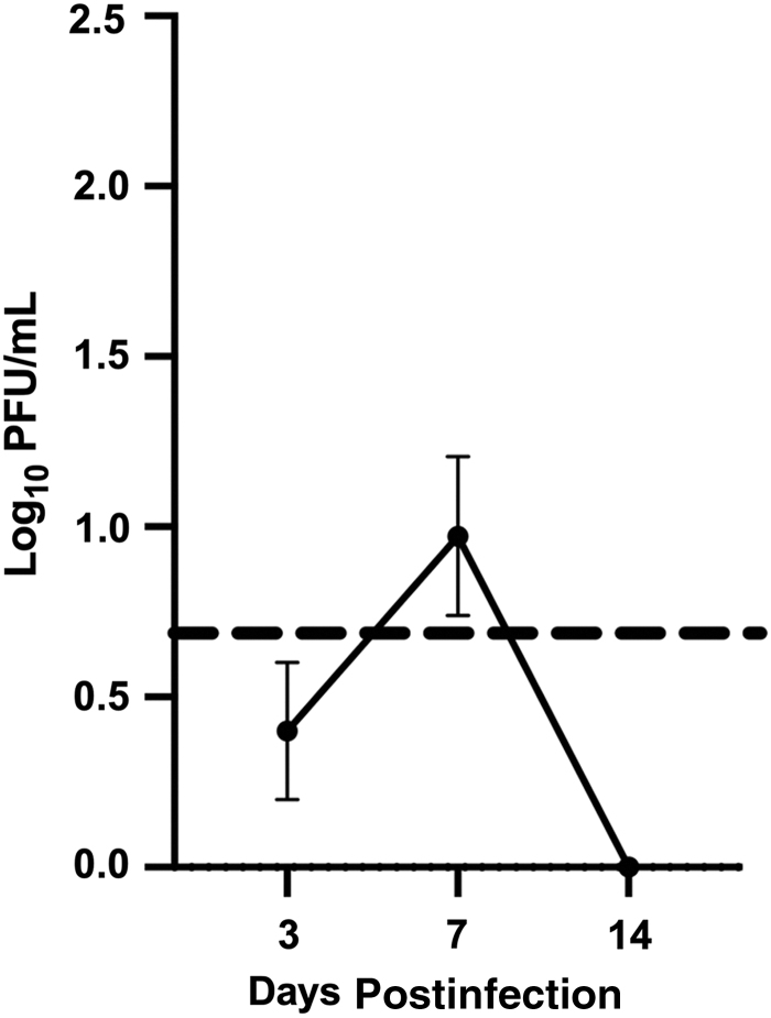
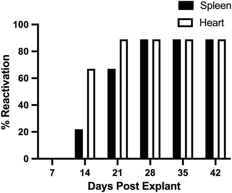
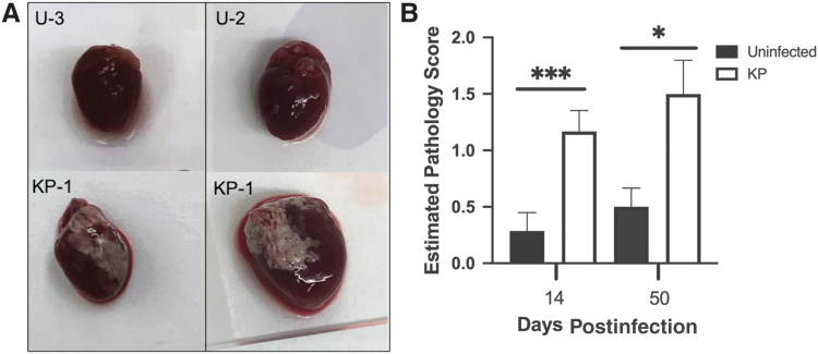
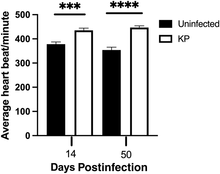
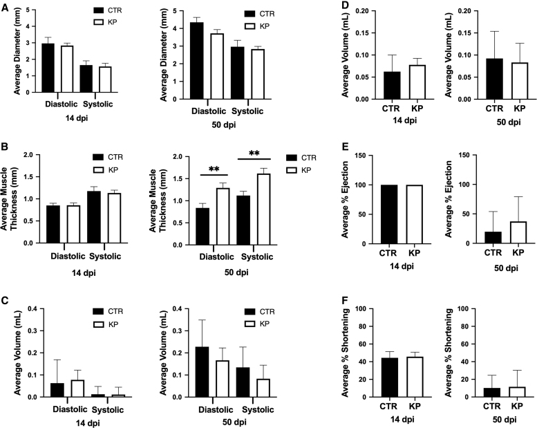
Human cytomegalovirus (HCMV) is associated with increased risk of chronic diseases of the heart and vasculature, including myocarditis, atherosclerosis, and transplant vasculopathy. To investigate CMV infection of the heart, murine cytomegalovirus (MCMV) was used to evaluate both acute and latent infection and the subsequent phenotypic and functional consequences of infection. Female BALB/c mice were intraperitoneally (i.p.) inoculated with 1 × 10 pfu of MCMV and evaluated at 14 and 50 days postinfection (dpi). At each time point, echocardiography was used to evaluate cardiac function and histology was conducted for phenotypic evaluation. MCMV replication in the heart was detected as early as 3 dpi and was no longer detectable at 14 dpi. Infected animals had significant cardiac pathology at 14 and 50 dpi when compared to uninfected controls. Histology revealed fibrosis of the heart as early as 14 dpi and the presence of white fibrous deposits on the surface of the heart. Functional evaluation showed significantly increased heart rate and muscle thickening in the latently infected animals when compared to the control animals. At 50 dpi, latent virus was measured by explant reactivation assay, demonstrating that MCMV establishes latency and is capable of reactivation from the heart, similar to other tissues such as spleen and salivary glands. Collectively, these studies illustrate that MCMV infection results in phenotypic alterations within the heart as early as 14 dpi, which progress to functional abnormalities during latency. These findings are similar to sinus tachycardia and hypertrophy of the heart muscle observed in cases of HCMV-induced acute myocarditis.
人巨细胞病毒(HCMV)与心脏和血管的慢性疾病风险增加有关,包括心肌炎、动脉粥样硬化和移植血管病。为了研究心脏的 CMV 感染,使用鼠巨细胞病毒(MCMV)来评估急性和潜伏感染以及随后感染的表型和功能后果。雌性 BALB/c 小鼠通过腹腔内(i.p.)接种 1×10 pfu 的 MCMV,并在感染后 14 和 50 天(dpi)进行评估。在每个时间点,使用超声心动图评估心脏功能,进行组织学评估以评估表型。早在 3dpi 就检测到心脏中的 MCMV 复制,在 14dpi 时不再可检测到。与未感染对照相比,感染动物在 14 和 50dpi 时具有明显的心脏病理学。组织学显示早在 14dpi 就出现心脏纤维化,并且心脏表面存在白色纤维沉积物。功能评估显示潜伏感染动物的心率和肌肉增厚明显高于对照动物。在 50dpi 时,通过外植体再激活测定测量潜伏病毒,表明 MCMV 建立潜伏并能够从心脏重新激活,类似于其他组织如脾脏和唾液腺。总之,这些研究表明,MCMV 感染早在 14dpi 就导致心脏内出现表型改变,在潜伏期间进展为功能异常。这些发现类似于 HCMV 诱导的急性心肌炎中观察到的窦性心动过速和心肌肥大。