TU Wien, Atominstitut, Stadionallee 2, Vienna, 1020, Austria.
Karlsruher Institute for Technology (KIT), ANKA Synchrotron Radiation Source, Hermann-von-Helmholtz-Platz 1, Eggenstein-Leopoldshafen, Karlsruhe, 76344, Germany.
Sci Rep. 2020 Apr 14;10(1):6301. doi: 10.1038/s41598-020-63325-9.
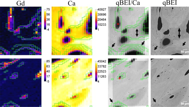
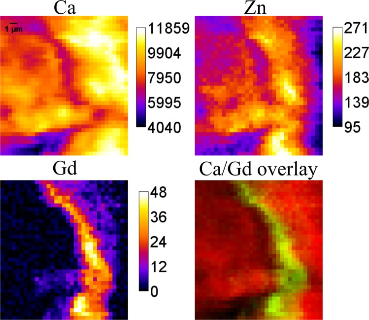
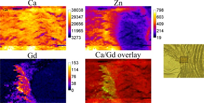
Gadolinium-based contrast agents (GBCAs) are frequently used in patients undergoing magnetic resonance imaging. In GBCAs gadolinium (Gd) is present in a bound chelated form. Gadolinium is a rare-earth element, which is normally not present in human body. Though the blood elimination half-life of contrast agents is about 90 minutes, recent studies demonstrated that some tissues retain gadolinium, which might further pose a health threat due to toxic effects of free gadolinium. It is known that the bone tissue can serve as a gadolinium depot, but so far only bulk measurements were performed. Here we present a summary of experiments in which for the first time we mapped gadolinium in bone biopsy from a male patient with idiopathic osteoporosis (without indication of renal impairment), who received MRI 8 months prior to biopsy. In our studies performed by means of synchrotron radiation induced micro- and submicro-X-ray fluorescence spectroscopy (SR-XRF), gadolinium was detected in human cortical bone tissue. The distribution of gadolinium displays a specific accumulation pattern. Correlation of elemental maps obtained at ANKA synchrotron with qBEI images (quantitative backscattered electron imaging) allowed assignment of Gd structures to the histological bone structures. Follow-up beamtimes at ESRF and Diamond Light Source using submicro-SR-XRF allowed resolving thin Gd structures in cortical bone, as well as correlating them with calcium and zinc.
钆基造影剂(GBCAs)常用于磁共振成像患者。在 GBCAs 中,钆(Gd)以结合螯合的形式存在。钆是一种稀土元素,在人体中通常不存在。尽管造影剂的血液消除半衰期约为 90 分钟,但最近的研究表明,一些组织保留了钆,由于游离钆的毒性作用,这可能进一步构成健康威胁。已知骨组织可以作为钆的储存库,但到目前为止,仅进行了批量测量。在这里,我们总结了实验,首次对一名患有特发性骨质疏松症(无肾功能损害迹象)的男性患者的骨活检进行了钆测绘,该患者在活检前 8 个月接受了 MRI。在我们通过同步辐射诱导的微区和亚微区 X 射线荧光光谱学(SR-XRF)进行的研究中,在人皮质骨组织中检测到了钆。钆的分布显示出特定的积累模式。在 ANKA 同步加速器上进行的元素图谱与 qBEI 图像(定量背散射电子成像)的相关性,允许将 Gd 结构分配到组织学骨结构上。在 ESRF 和 Diamond Light Source 上使用亚微 SR-XRF 进行的后续束流时间允许解析皮质骨中的薄 Gd 结构,并将其与钙和锌相关联。