Department of Radiology, Nagoya University Graduate School of Medicine.
Department of Otorhinolaryngology, Nagoya University Graduate School of Medicine.
Magn Reson Med Sci. 2021 Mar 1;20(1):91-98. doi: 10.2463/mrms.mp.2020-0030. Epub 2020 Apr 15.
To evaluate the feasibility for the detection of slight contrast effects after intravenous administration of single dose gadolinium-based contrast agent (IV-SD-GBCA), the time course of the GBCA distribution up to 24 h was examined in various fluid spaces and brain parenchyma using 3D-real IR imaging and MR fingerprinting (MRF).
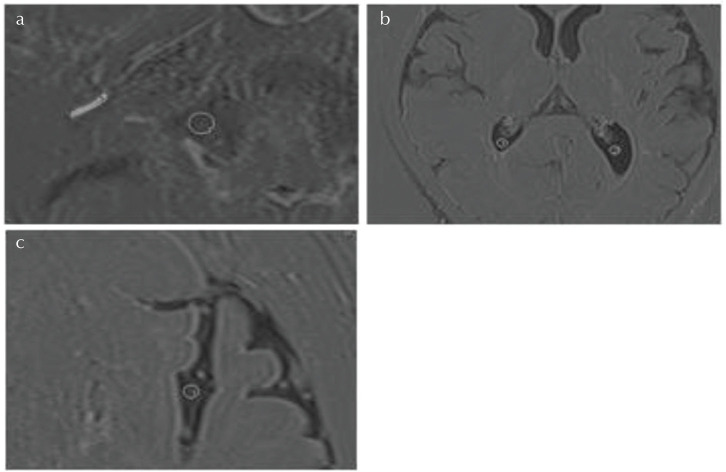
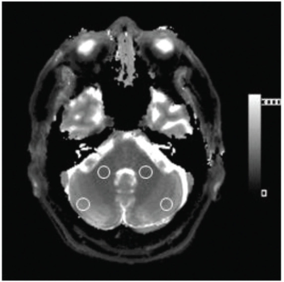
Twenty-four patients with a suspicion of endolymphatic hydrops were scanned at pre-administration and at 10 min, 4 and 24 h post-IV-SD-GBCA. 3D-real IR images and MRF at the level of the internal auditory canal were obtained. The signal intensity on the 3D-real IR image of the cerebrospinal fluid (CSF) in the cerebellopontine angle cistern (CPA), Sylvian fissure (Syl), lateral ventricle (LV), and cochlear perilymph (CPL) was measured. The T and T values of cerebellar gray (GM) and white matter (WM) were measured using MRF. Each averaged value at the various time points was compared using an analysis of variance.
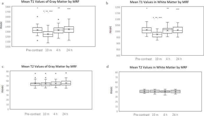
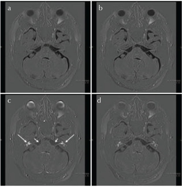
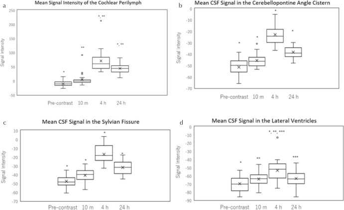
The signal intensity on the 3D-real IR image in each CSF region peaked at 4 h, and was decreased significantly by 24 h (P< 0.05). All patients had a maximum signal intensity at 4 h in the CPA, and Syl. The mean CPL signal intensity peaked at 4 h and decreased significantly by 24 h (P < 0.05). All patients but two had a maximum signal intensity at 4 h. Regarding the T value in the cerebellar WM and GM, the T value at 10 min post-IV-GBCA was significantly decreased compared to the pre-contrast scan, but no significant difference was observed at the other time points. There was no significant change in T in the gray or white matter at any of the time points.
Time course of GBCA after IV-SD-GBCA could be evaluated by 3D-real IR imaging in CSF spaces and in the brain by MRF.
评估单次静脉注射钆基对比剂(IV-SD-GBCA)后检测轻微对比效果的可行性,使用 3D 真实反转成像和磁共振指纹识别(MRF)检查 GBCA 在 24 小时内分布至各种体液空间和脑实质的时间过程。
24 例怀疑内淋巴积水的患者在给药前和给药后 10 分钟、4 小时和 24 小时进行扫描。获得内听道水平的 3D 真实反转图像和 MRF。测量桥小脑角池(CPA)、大脑外侧裂(Syl)、侧脑室(LV)和耳蜗外淋巴(CPL)的脑脊液(CSF)在 3D 真实反转图像上的信号强度。使用 MRF 测量小脑灰质(GM)和白质(WM)的 T 和 T 值。使用方差分析比较各个时间点的每个平均值。
各 CSF 区 3D 真实反转图像的信号强度在 4 小时达到峰值,24 小时时显著降低(P<0.05)。所有患者在 CPA 和 Syl 均在 4 小时达到最大信号强度。CPL 的平均信号强度在 4 小时达到峰值,并在 24 小时时显著降低(P<0.05)。除两名患者外,所有患者均在 4 小时达到最大信号强度。关于小脑 WM 和 GM 的 T 值,IV-GBCA 后 10 分钟的 T 值与对比前扫描相比显著降低,但在其他时间点无显著差异。在任何时间点,GM 和 WM 的 T 值均无显著变化。
通过 MRF 可以在 CSF 空间和脑实质中使用 3D 真实反转成像评估 IV-SD-GBCA 后 GBCA 的时间过程。