Jung Yoon Gyo, Jung Sang Ku, Lee Byung Jou, Lee Subum, Jeong Seong Kyun, Kim Myeongjong, Park Jin Hoon
Department of Neurosurgery, Asan Medical Center, University of Ulsan College of Medicine.
Department of Emergency Medicine, Gangneung Asan Hospital, University of Ulsan College of Medicine.
Neurol Med Chir (Tokyo). 2020 May 15;60(5):231-243. doi: 10.2176/nmc.ra.2019-0189. Epub 2020 Apr 15.
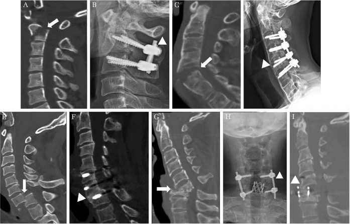
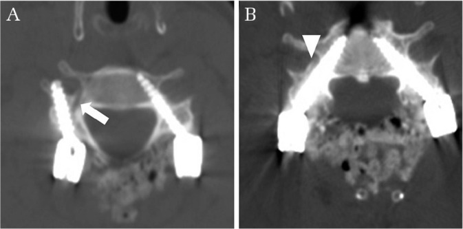
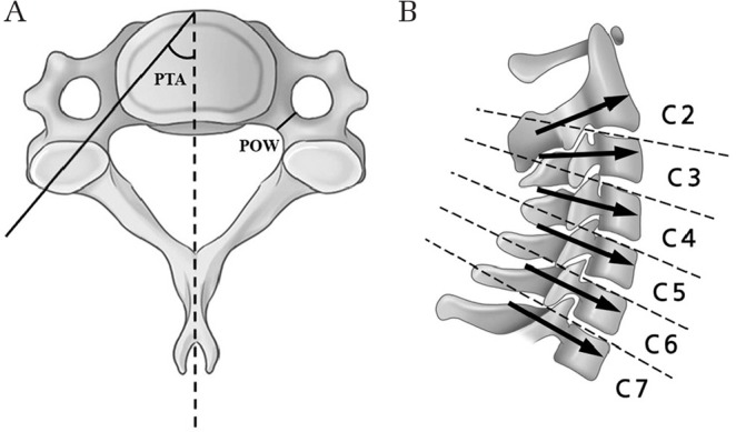
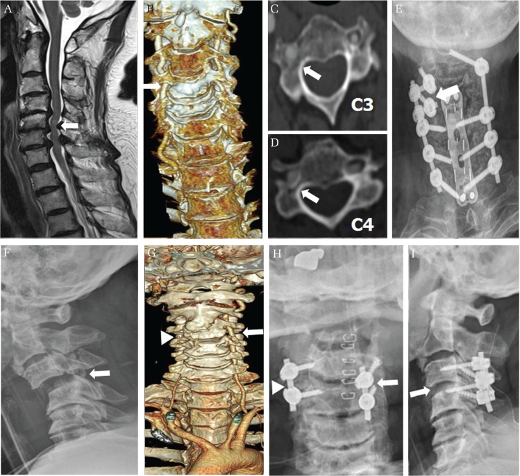
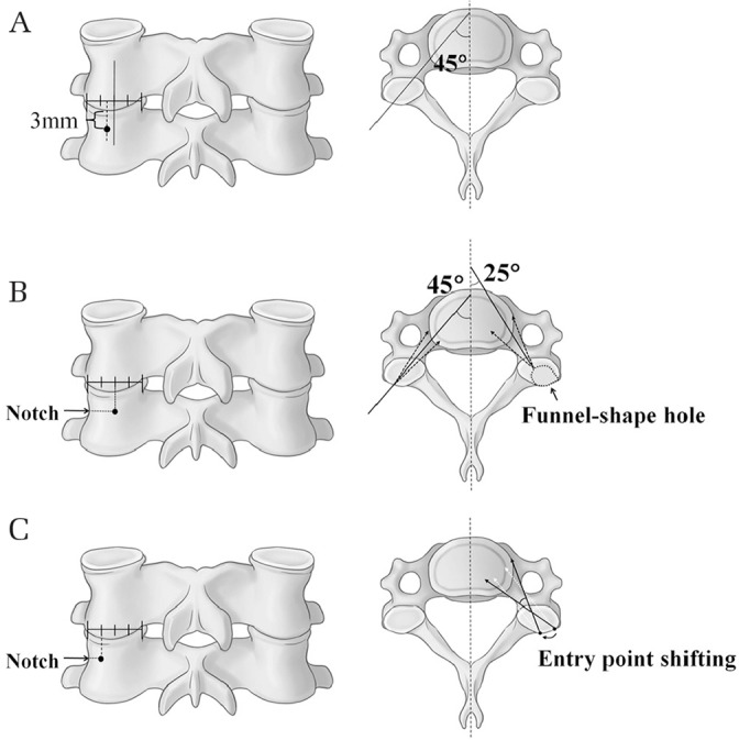
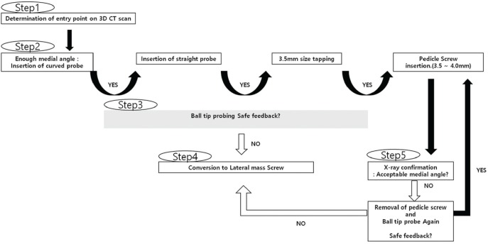
This study aimed to review information on the subaxial cervical pedicle screw (CPS) including recent anatomical considerations, entry points, placement techniques, accuracy, learning curve, and complications. Relevant literatures were reviewed, and the authors' experiences were summarized. The CPS is used for reconstruction of unstable cervical spine and achieves superior biomechanical stability compared to other fixation techniques. Various insertion and guidance techniques are established, among which, lateral fluoroscopy-assisted placement is the most common and cost-effective technique. Generally, placement under imaging guidance is more accurate than other techniques, and a three-dimensional template allows optimal trajectory for each pedicle regardless of intraoperative changes in spinal alignment. The free-hand technique using a curved pedicle probe without a funnel-like hole increases screw stability and reduces operation time, radiation exposure, and soft tissue injury. Compared to conventional lateral fluoroscopy-assisted placement, free-hand CPS placement by trained surgeons achieves superior accuracy comparable to that of image-guided navigation; in general, 30 training cases are sufficient for learning a safe and accurate technique for CPS placement. The complications of subaxial CPS are classified into three categories: complications due to screw misplacement, complications without screw misplacement, and others. Inexperienced surgeons may benefit from advanced techniques; however, the accuracy of CPS ultimately depends on the surgeon's experience. Inexperienced surgeons should master the placement of the thoracolumbar pedicle screw in real practice and practice CPS insertion using cadavers. During the initial phase of the learning curve, careful preparation of surgery, reiterated identification, patterned safety steps, and supervision of the expert are necessary.
本研究旨在回顾关于下颈椎椎弓根螺钉(CPS)的信息,包括近期的解剖学考量、进针点、置入技术、准确性、学习曲线及并发症。回顾了相关文献,并总结了作者的经验。CPS用于不稳定颈椎的重建,与其他固定技术相比,能实现更好的生物力学稳定性。已建立了多种置入和引导技术,其中,侧位透视辅助置入是最常用且性价比最高的技术。一般来说,在影像引导下置入比其他技术更准确,三维模板可为每个椎弓根提供最佳轨迹,而不受术中脊柱对线变化的影响。使用无漏斗状孔的弯形椎弓根探子的徒手技术可增加螺钉稳定性,减少手术时间、辐射暴露及软组织损伤。与传统侧位透视辅助置入相比,经培训的外科医生进行徒手CPS置入可达到与影像引导导航相当的更高准确性;一般而言,30例培训病例足以学会安全准确的CPS置入技术。下颈椎CPS的并发症分为三类:螺钉误置引起的并发症、无螺钉误置的并发症及其他并发症。经验不足的外科医生可能会从先进技术中受益;然而,CPS的准确性最终取决于外科医生的经验。经验不足的外科医生应在实际操作中掌握胸腰椎椎弓根螺钉的置入,并使用尸体练习CPS置入。在学习曲线的初始阶段,精心准备手术、反复确认、制定安全步骤及专家监督是必要的。