• 文献检索
  • 文档翻译
  • 深度研究
  • 学术资讯
  • Suppr Zotero 插件Zotero 插件
  • 邀请有礼
  • 套餐&价格
  • 历史记录
应用&插件
Suppr Zotero 插件Zotero 插件浏览器插件Mac 客户端Windows 客户端微信小程序
定价
高级版会员购买积分包购买API积分包
服务
文献检索文档翻译深度研究API 文档MCP 服务
关于我们
关于 Suppr公司介绍联系我们用户协议隐私条款
关注我们

Suppr 超能文献

核心技术专利:CN118964589B侵权必究
粤ICP备2023148730 号-1Suppr @ 2026

文献检索

告别复杂PubMed语法,用中文像聊天一样搜索,搜遍4000万医学文献。AI智能推荐,让科研检索更轻松。

立即免费搜索

文件翻译

保留排版,准确专业,支持PDF/Word/PPT等文件格式,支持 12+语言互译。

免费翻译文档

深度研究

AI帮你快速写综述,25分钟生成高质量综述,智能提取关键信息,辅助科研写作。

立即免费体验

描述人类周围神经退化的细胞和分子特征。

Characterising cellular and molecular features of human peripheral nerve degeneration.

机构信息

Peripheral Nerve Injury Research Unit, Royal National Orthopaedic Hospital, Stanmore, UK.

Department of Pharmacology, UCL School of Pharmacy, University College London, London, WC1N 1AX, UK.

出版信息

Acta Neuropathol Commun. 2020 Apr 17;8(1):51. doi: 10.1186/s40478-020-00921-w.

DOI:10.1186/s40478-020-00921-w
PMID:32303273
原文链接:https://pmc.ncbi.nlm.nih.gov/articles/PMC7164159/
Abstract

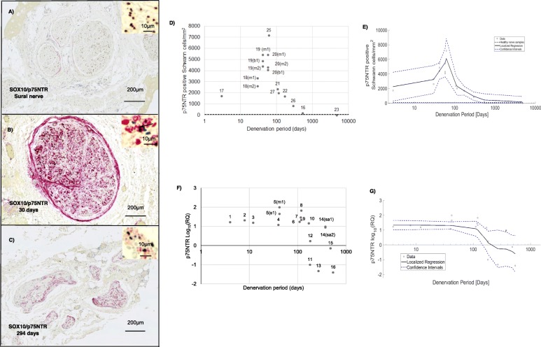
Nerve regeneration is a key biological process in those recovering from neural trauma. From animal models it is known that the regenerative capacity of the peripheral nervous system (PNS) relies heavily on the remarkable ability of Schwann cells to undergo a phenotypic shift from a myelinating phenotype to one that is supportive of neural regeneration. In rodents, a great deal is known about the molecules that control this process, such as the transcription factors c-Jun and early growth response protein 2 (EGR2/KROX20), or mark the cells and cellular changes involved, including SOX10 and P75 neurotrophin receptor (p75NTR). However, ethical and practical challenges associated with studying human nerve injury have meant that little is known about human nerve regeneration.The present study addresses this issue, analysing 34 denervated and five healthy nerve samples from 27 patients retrieved during reconstructive nerve procedures. Using immunohistochemistry and Real-Time quantitative Polymerase Chain Reaction (RT-qPCR), the expression of SOX10, c-Jun, p75NTR and EGR2 was assessed in denervated samples and compared to healthy nerve. Nonparametric smoothing linear regression was implemented to better visualise trends in the expression of these markers across denervated samples.It was found, first, that two major genes associated with repair Schwann cells in rodents, c-Jun and p75NTR, are also up-regulated in acutely injured human nerves, while the myelin associated transcription factor EGR2 is down-regulated, observations that encourage the view that rodent models are relevant for learning about human nerve injury. Second, as in rodents, the expression of c-Jun and p75NTR declines during long-term denervation. In rodents, diminishing c-Jun and p75NTR levels mark the general deterioration of repair cells during chronic denervation, a process thought to be a major obstacle to effective nerve repair. The down-regulation of c-Jun and p75NTR reported here provides the first molecular evidence that also in humans, repair cells deteriorate during chronic denervation.

摘要

神经再生是神经创伤后恢复的关键生物学过程。从动物模型可知,周围神经系统(PNS)的再生能力在很大程度上依赖于施万细胞经历表型转变的显著能力,从髓鞘形成表型转变为支持神经再生的表型。在啮齿动物中,人们对控制这一过程的分子有了很多了解,例如转录因子 c-Jun 和早期生长反应蛋白 2(EGR2/KROX20),或者标记涉及的细胞和细胞变化,包括 SOX10 和 P75 神经营养因子受体(p75NTR)。然而,与研究人类神经损伤相关的伦理和实际挑战意味着,人们对人类神经再生知之甚少。本研究解决了这个问题,分析了 27 名患者在重建性神经手术中获取的 34 个去神经支配和 5 个健康神经样本。通过免疫组织化学和实时定量聚合酶链反应(RT-qPCR),在去神经支配样本中评估了 SOX10、c-Jun、p75NTR 和 EGR2 的表达,并与健康神经进行了比较。实施了非参数平滑线性回归,以更好地观察这些标记物在去神经支配样本中的表达趋势。结果发现,首先,与修复 Schwann 细胞相关的两种主要基因在啮齿动物中,c-Jun 和 p75NTR,也在急性损伤的人类神经中上调,而与髓鞘相关的转录因子 EGR2 下调,这些观察结果鼓励人们认为啮齿动物模型与学习人类神经损伤有关。其次,与啮齿动物一样,c-Jun 和 p75NTR 的表达在长期去神经支配期间下降。在啮齿动物中,c-Jun 和 p75NTR 水平的降低标志着修复细胞在慢性去神经支配过程中的普遍恶化,这一过程被认为是有效神经修复的主要障碍。这里报道的 c-Jun 和 p75NTR 的下调提供了第一个分子证据,表明在人类中,修复细胞在慢性去神经支配过程中也会恶化。

https://cdn.ncbi.nlm.nih.gov/pmc/blobs/a66f/7164159/42f7b8d6efc5/40478_2020_921_Fig6_HTML.jpg
https://cdn.ncbi.nlm.nih.gov/pmc/blobs/a66f/7164159/32739a21c4b5/40478_2020_921_Fig1_HTML.jpg
https://cdn.ncbi.nlm.nih.gov/pmc/blobs/a66f/7164159/350655fe7412/40478_2020_921_Fig2_HTML.jpg
https://cdn.ncbi.nlm.nih.gov/pmc/blobs/a66f/7164159/a27cdfe7137e/40478_2020_921_Fig3_HTML.jpg
https://cdn.ncbi.nlm.nih.gov/pmc/blobs/a66f/7164159/5af0f29d058c/40478_2020_921_Fig4_HTML.jpg
https://cdn.ncbi.nlm.nih.gov/pmc/blobs/a66f/7164159/6ff0bf0fc369/40478_2020_921_Fig5_HTML.jpg
https://cdn.ncbi.nlm.nih.gov/pmc/blobs/a66f/7164159/42f7b8d6efc5/40478_2020_921_Fig6_HTML.jpg

相似文献

1
Characterising cellular and molecular features of human peripheral nerve degeneration.描述人类周围神经退化的细胞和分子特征。
Acta Neuropathol Commun. 2020 Apr 17;8(1):51. doi: 10.1186/s40478-020-00921-w.
2
Neurotrophin-3 promotes peripheral nerve regeneration by maintaining a repair state of Schwann cells after chronic denervation via the TrkC/ERK/c-Jun pathway.神经营养因子-3 通过 TrkC/ERK/c-Jun 通路在慢性去神经支配后维持施万细胞的修复状态,从而促进周围神经再生。
J Transl Med. 2023 Oct 17;21(1):733. doi: 10.1186/s12967-023-04609-2.
3
Chronically denervated rat Schwann cells respond to GGF in vitro.慢性去神经支配的大鼠雪旺细胞在体外对神经生长因子有反应。
Glia. 1998 Nov;24(3):290-303.
4
Dynamic regulation of Schwann cell enhancers after peripheral nerve injury.外周神经损伤后雪旺细胞增强子的动态调控
J Biol Chem. 2015 Mar 13;290(11):6937-50. doi: 10.1074/jbc.M114.622878. Epub 2015 Jan 22.
5
mTORC1 Is Transiently Reactivated in Injured Nerves to Promote c-Jun Elevation and Schwann Cell Dedifferentiation.mTORC1 在受损神经中短暂激活以促进 c-Jun 升高和施万细胞去分化。
J Neurosci. 2018 May 16;38(20):4811-4828. doi: 10.1523/JNEUROSCI.3619-17.2018. Epub 2018 Apr 25.
6
Contribution of Schwann Cells to Remyelination in a Naturally Occurring Canine Model of CNS Neuroinflammation.在中枢神经系统神经炎症的自然发生犬模型中施万细胞对再髓鞘化的作用
PLoS One. 2015 Jul 21;10(7):e0133916. doi: 10.1371/journal.pone.0133916. eCollection 2015.
7
Graded Elevation of c-Jun in Schwann Cells : Gene Dosage Determines Effects on Development, Remyelination, Tumorigenesis, and Hypomyelination.雪旺细胞中c-Jun的分级上调:基因剂量决定对发育、髓鞘再生、肿瘤发生和髓鞘形成不足的影响
J Neurosci. 2017 Dec 13;37(50):12297-12313. doi: 10.1523/JNEUROSCI.0986-17.2017. Epub 2017 Nov 6.
8
Schwann cell p75NTR prevents spontaneous sensory reinnervation of the adult spinal cord.施万细胞 p75NTR 防止成年脊髓的自发性感觉神经再支配。
Brain. 2010 Feb;133(Pt 2):421-32. doi: 10.1093/brain/awp316. Epub 2010 Jan 3.
9
Adult skin-derived precursor Schwann cells exhibit superior myelination and regeneration supportive properties compared to chronically denervated nerve-derived Schwann cells.与慢性去神经支配的神经源性施万细胞相比,成人皮肤源性前体细胞施万细胞表现出更好的髓鞘形成和再生支持特性。
Exp Neurol. 2016 Apr;278:127-42. doi: 10.1016/j.expneurol.2016.02.006. Epub 2016 Feb 6.
10
Failures of nerve regeneration caused by aging or chronic denervation are rescued by restoring Schwann cell c-Jun.衰老或慢性去神经支配引起的神经再生失败可通过恢复许旺细胞 c-Jun 来挽救。
Elife. 2021 Jan 21;10:e62232. doi: 10.7554/eLife.62232.

引用本文的文献

1
The Neuroregenerative Effects of IncobotulinumtoxinA (Inco/A) in a Nerve Lesion Model of the Rat.因可保妥适(Inco/A)在大鼠神经损伤模型中的神经再生作用
Int J Mol Sci. 2025 Aug 2;26(15):7482. doi: 10.3390/ijms26157482.
2
Roles of Peripheral Nerves in Tumor Initiation and Progression.外周神经在肿瘤起始和进展中的作用。
Int J Mol Sci. 2025 Jul 22;26(15):7064. doi: 10.3390/ijms26157064.
3
Axonal regeneration and innervation ratio following supercharged end-to-side nerve transfer.强化端侧神经转位后的轴突再生与支配率

本文引用的文献

1
Peripheral nerve injury induced changes in the spinal cord and strategies to counteract/enhance the changes to promote nerve regeneration.周围神经损伤引起脊髓的变化以及对抗/增强这些变化以促进神经再生的策略。
Neural Regen Res. 2020 Feb;15(2):189-198. doi: 10.4103/1673-5374.265540.
2
Peripheral Nerve Regeneration Is Independent From Schwann Cell p75 Expression.周围神经再生独立于雪旺细胞p75的表达。
Front Cell Neurosci. 2019 May 29;13:235. doi: 10.3389/fncel.2019.00235. eCollection 2019.
3
The Effects of Surgical Antiseptics and Time Delays on RNA Isolated From Human and Rodent Peripheral Nerves.
Front Cell Dev Biol. 2025 Feb 12;13:1513321. doi: 10.3389/fcell.2025.1513321. eCollection 2025.
4
Refinement of Protein Extraction Protocols for Human Peripheral Nerve Tissue.人类外周神经组织蛋白质提取方案的优化
ACS Omega. 2025 Feb 2;10(5):5111-5118. doi: 10.1021/acsomega.4c11373. eCollection 2025 Feb 11.
5
The role and mechanism of Schwann cells in the repair of peripheral nerve injury.施万细胞在周围神经损伤修复中的作用及机制。
Cell Tissue Res. 2025 Apr;400(1):81-95. doi: 10.1007/s00441-025-03957-3. Epub 2025 Feb 15.
6
Effects of PEMF and LIPUS Therapy on the Expression of Genes Related to Peripheral Nerve Regeneration in Schwann Cells.脉冲电磁场和低强度脉冲超声疗法对雪旺细胞中与周围神经再生相关基因表达的影响。
Int J Mol Sci. 2024 Nov 28;25(23):12791. doi: 10.3390/ijms252312791.
7
Interpretation of Data from Translational Rodent Nerve Injury and Repair Models.转化型啮齿动物神经损伤与修复模型的数据解读。
Hand Clin. 2024 Aug;40(3):429-440. doi: 10.1016/j.hcl.2024.03.004. Epub 2024 May 19.
8
Influence of Magnesium Degradation on Schwannoma Cell Responses to Nerve Injury Using an In Vitro Injury Model.使用体外损伤模型研究镁降解对雪旺瘤细胞对神经损伤反应的影响。
J Funct Biomater. 2024 Mar 31;15(4):88. doi: 10.3390/jfb15040088.
9
Anisotropic microtopography surface of chitosan scaffold regulating skin precursor-derived Schwann cells towards repair phenotype promotes neural regeneration.壳聚糖支架的各向异性微形貌表面将皮肤前体来源的雪旺细胞调节为修复表型,从而促进神经再生。
Regen Biomater. 2024 Jan 27;11:rbae005. doi: 10.1093/rb/rbae005. eCollection 2024.
10
The Dynamics of Nerve Degeneration and Regeneration in a Healthy Milieu and in Diabetes.在健康环境和糖尿病中神经变性和再生的动态。
Int J Mol Sci. 2023 Oct 16;24(20):15241. doi: 10.3390/ijms242015241.
手术消毒剂和时间延迟对从人和啮齿动物外周神经分离的RNA的影响。
Front Cell Neurosci. 2019 May 22;13:189. doi: 10.3389/fncel.2019.00189. eCollection 2019.
4
Role of macrophages in peripheral nerve injury and repair.巨噬细胞在周围神经损伤与修复中的作用。
Neural Regen Res. 2019 Aug;14(8):1335-1342. doi: 10.4103/1673-5374.253510.
5
The Success and Failure of the Schwann Cell Response to Nerve Injury.施万细胞对神经损伤反应的成功与失败
Front Cell Neurosci. 2019 Feb 11;13:33. doi: 10.3389/fncel.2019.00033. eCollection 2019.
6
Nerve transfers for peripheral nerve injury in the upper limb: a case-based review.上肢周围神经损伤的神经转移:基于病例的回顾。
Bone Joint J. 2019 Feb;101-B(2):124-131. doi: 10.1302/0301-620X.101B2.BJJ-2018-0839.R1.
7
Repair Schwann cell update: Adaptive reprogramming, EMT, and stemness in regenerating nerves.修复雪旺细胞更新:再生神经中的适应性重编程、上皮间质转化和干性。
Glia. 2019 Mar;67(3):421-437. doi: 10.1002/glia.23532. Epub 2019 Jan 11.
8
ImageJ2: ImageJ for the next generation of scientific image data.ImageJ2:面向下一代科学图像数据的ImageJ。
BMC Bioinformatics. 2017 Nov 29;18(1):529. doi: 10.1186/s12859-017-1934-z.
9
Aging Schwann cells: mechanisms, implications, future directions.衰老雪旺细胞:机制、意义、未来方向。
Curr Opin Neurobiol. 2017 Dec;47:203-208. doi: 10.1016/j.conb.2017.10.022. Epub 2017 Nov 21.
10
After Nerve Injury, Lineage Tracing Shows That Myelin and Remak Schwann Cells Elongate Extensively and Branch to Form Repair Schwann Cells, Which Shorten Radically on Remyelination.神经损伤后,谱系追踪显示髓鞘和雷马克雪旺细胞会广泛伸长并分支形成修复性雪旺细胞,而这些修复性雪旺细胞在髓鞘再生时会显著缩短。
J Neurosci. 2017 Sep 13;37(37):9086-9099. doi: 10.1523/JNEUROSCI.1453-17.2017. Epub 2017 Aug 3.