Zhu Qingyun, Guo Yuehui, Chen Shiwei, Fu Daiquan, Li Yanxiang, Li Zhi, Ni Caifang
Department of Interventional Radiology, The First Affiliated Hospital of Soochow University, Suzhou 215006, People's Republic of China.
Department of Intervention, Gongli Hospital of Shanghai Pudong New Area, Shanghai 200135, People's Republic of China.
Onco Targets Ther. 2020 Apr 2;13:2807-2817. doi: 10.2147/OTT.S240803. eCollection 2020.
Irinotecan (IRI) is considered an option for second-line treatment of advanced gastric cancer; however, acquired drug resistance currently limits its clinical application. Recently, many researchers have shown that autophagy plays a crucial role in the resistance of tumor cells to chemotherapy and radiotherapy. In this study, we investigated the relationship between autophagy and antitumor activity of IRI in gastric cancer cells.
We used MTT assay, flow cytometry and immunofluorescence staining to detect viability, apoptosis and autophagy in gastric cancer. Western blotting assay was used to determine the expression of LC3, Beclin-1, P62, cleaved PARP and Caspase 3. In vivo animal study was performed finally.
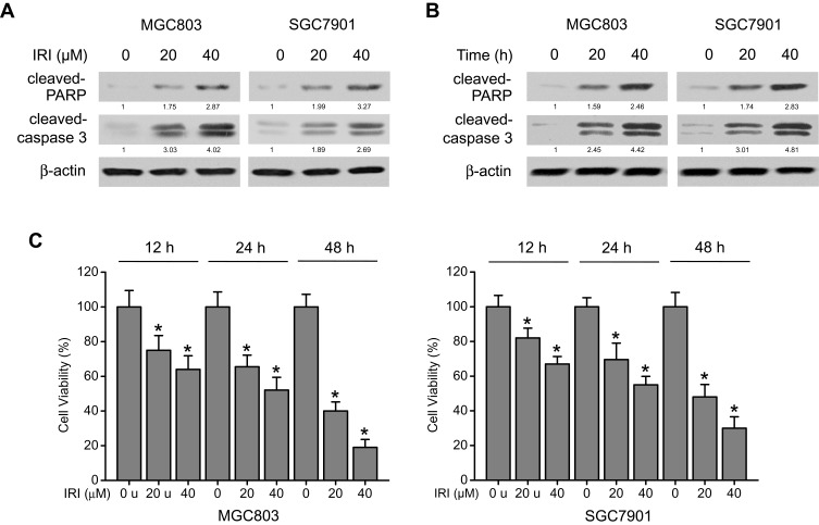
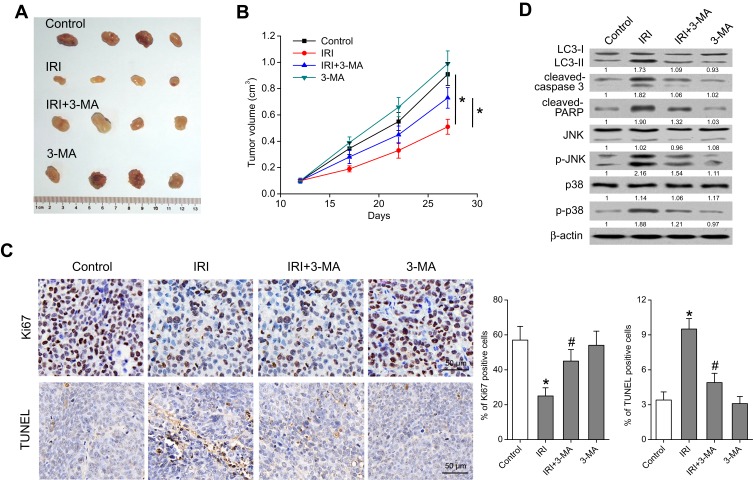
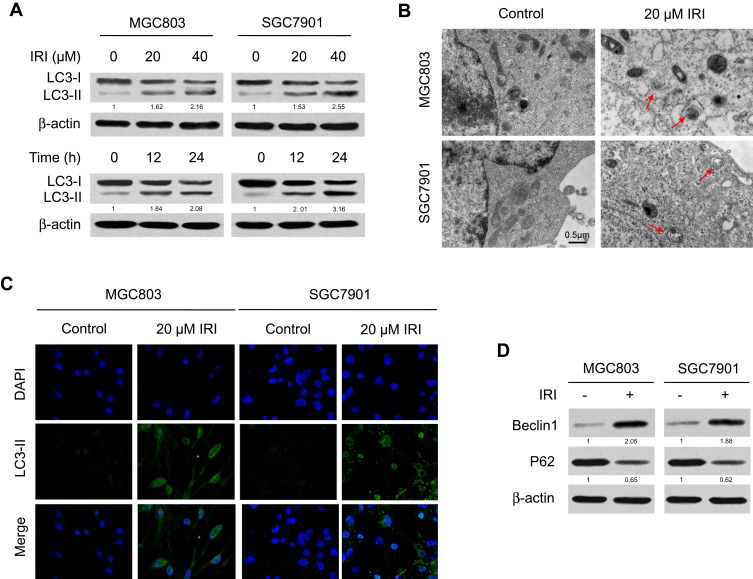
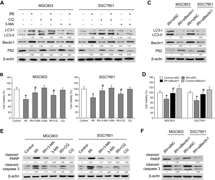
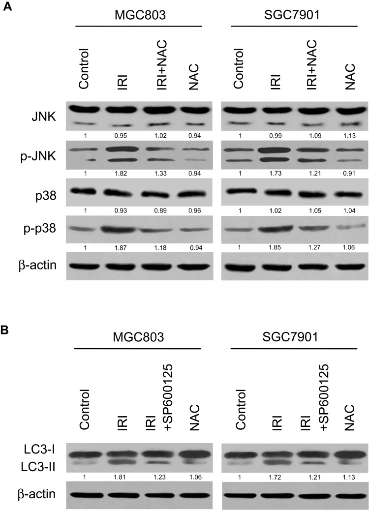
We found that IRI treatment dose- and time-dependently inhibited growth and induced apoptosis in gastric cancer cells. Moreover, IRI treatment caused autophagy in these cells, whereas autophagy inhibitors-3-methyladenine (3-MA), chloroquine (CQ), and Beclin-1 small interfering RNA (siRNA)-suppressed cytotoxicity of IRI. A mechanistic analysis showed that IRI-induced autophagy and apoptosis were related to increased reactive oxygen species (ROS) accumulation and activation of the JNK- and p38-MAPK pathways. Further in vivo experiments revealed that IRI suppressed tumor growth, induced autophagy, and stimulated the JNK- and p38-MAPK pathways, whereas 3-MA attenuated these effects.
Taken together, these results indicate that IRI stimulates the ROS-related JNK- and p38-MAPK pathways to promote autophagy-dependent apoptosis. Thus, a combination of IRI with a pharmacological autophagy enhancer may be a promising therapeutic strategy against gastric cancer.
伊立替康(IRI)被认为是晚期胃癌二线治疗的一种选择;然而,获得性耐药目前限制了其临床应用。最近,许多研究人员表明自噬在肿瘤细胞对化疗和放疗的耐药性中起关键作用。在本研究中,我们调查了自噬与IRI在胃癌细胞中的抗肿瘤活性之间的关系。
我们使用MTT法、流式细胞术和免疫荧光染色来检测胃癌中的细胞活力、凋亡和自噬。采用蛋白质免疫印迹法测定LC3、Beclin-1、P62、裂解的PARP和Caspase 3的表达。最后进行体内动物研究。
我们发现IRI处理剂量和时间依赖性地抑制胃癌细胞生长并诱导凋亡。此外,IRI处理导致这些细胞发生自噬,而自噬抑制剂3-甲基腺嘌呤(3-MA)、氯喹(CQ)和Beclin-1小干扰RNA(siRNA)抑制了IRI的细胞毒性。机制分析表明,IRI诱导的自噬和凋亡与活性氧(ROS)积累增加以及JNK和p38丝裂原活化蛋白激酶(MAPK)途径的激活有关。进一步的体内实验表明,IRI抑制肿瘤生长,诱导自噬,并刺激JNK和p38 MAPK途径,而3-MA减弱了这些作用。
综上所述,这些结果表明IRI刺激ROS相关的JNK和p38 MAPK途径以促进自噬依赖性凋亡。因此,IRI与药理学自噬增强剂联合使用可能是一种有前景的胃癌治疗策略。