Department of Otolaryngology-Head and Neck Surgery, Massachusetts Eye and Ear Infirmary, Boston, Massachusetts, USA.
Department of Otolaryngology-Head and Neck Surgery, Harvard Medical School, Boston, Massachusetts, USA.
Hum Brain Mapp. 2020 Aug 15;41(12):3253-3265. doi: 10.1002/hbm.25012. Epub 2020 Apr 20.
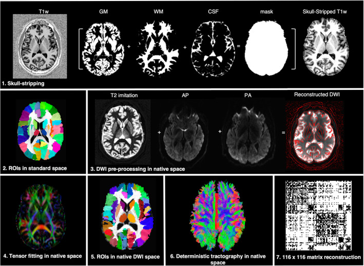
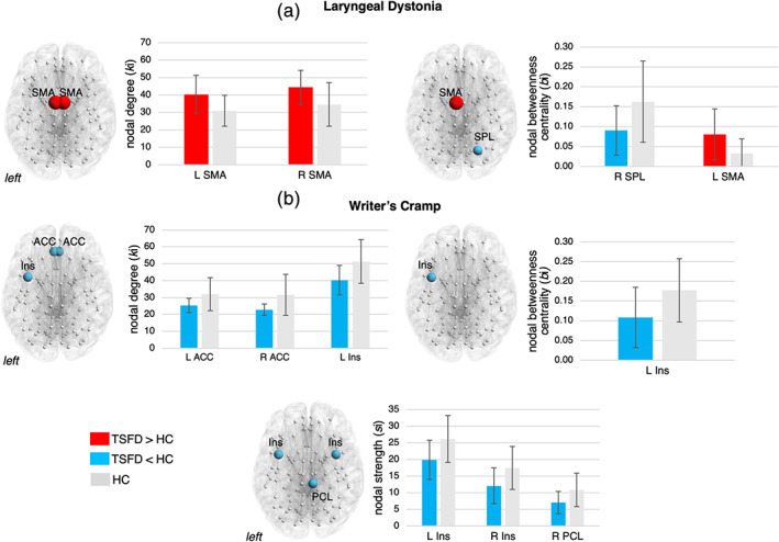
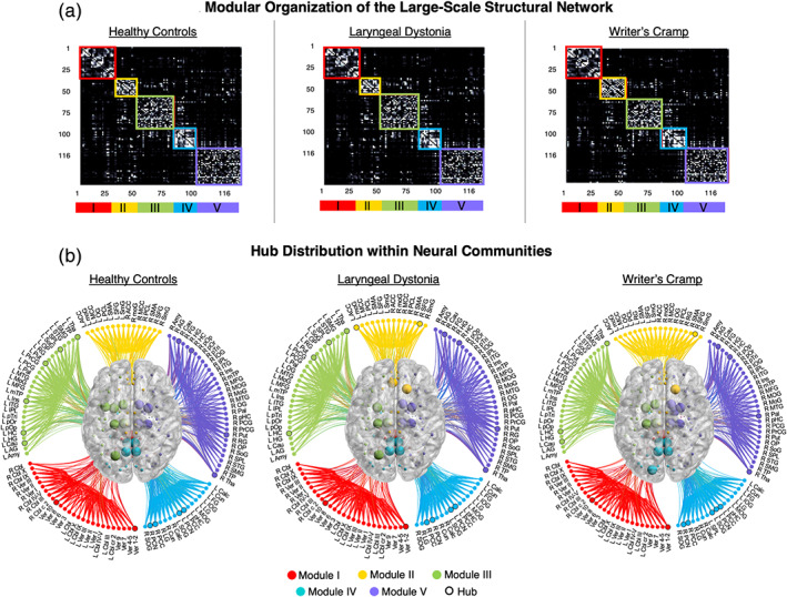
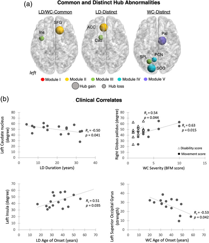
The emerging view of dystonia is that of a large-scale functional network disorder, in which the communication is disrupted between sensorimotor cortical areas, basal ganglia, thalamus, and cerebellum. The structural underpinnings of functional alterations in dystonia are, however, poorly understood. Notably, it is unclear whether structural changes form a larger-scale dystonic network or rather remain focal to isolated brain regions, merely underlying their functional abnormalities. Using diffusion-weighted imaging and graph theoretical analysis, we examined inter-regional white matter connectivity of the whole-brain structural network in two different forms of task-specific focal dystonia, writer's cramp and laryngeal dystonia, compared to healthy individuals. We show that, in addition to profoundly altered functional network in focal dystonia, its structural connectome is characterized by large-scale aberrations due to abnormal transfer of prefrontal and parietal nodes between neural communities and the reorganization of normal hub architecture, commonly involving the insula and superior frontal gyrus in patients compared to controls. Other prominent common changes involved the basal ganglia, parietal and cingulate cortical regions, whereas premotor and occipital abnormalities distinctly characterized the two forms of dystonia. We propose a revised pathophysiological model of focal dystonia as a disorder of both functional and structural connectomes, where dystonia form-specific abnormalities underlie the divergent mechanisms in the development of distinct clinical symptomatology. These findings may guide the development of novel therapeutic strategies directed at targeted neuromodulation of pathophysiological brain regions for the restoration of their structural and functional connectivity.
扭转痉挛的新观点是一种大规模的功能网络障碍,在这种障碍中,感觉运动皮质区、基底节、丘脑和小脑之间的通讯被中断。然而,扭转痉挛中功能改变的结构基础理解甚少。值得注意的是,尚不清楚结构变化是否形成更大规模的扭转网络,还是仍然局限于孤立的脑区,仅仅是其功能异常的基础。我们使用弥散加权成像和图论分析,比较了两种不同形式的任务特异性局灶性肌张力障碍(书写痉挛和喉痉挛)与健康个体之间全脑结构网络的区域间白质连接。我们表明,除了局灶性肌张力障碍中明显改变的功能网络外,其结构连接组还表现出大规模异常,这是由于前额叶和顶叶节点在神经群之间的异常转移以及正常枢纽结构的重组导致的,与对照组相比,患者中常见的异常涉及岛叶和额上回。其他突出的共同变化涉及基底节、顶叶和扣带回皮质区域,而运动前区和枕叶的异常则明显特征化了两种形式的肌张力障碍。我们提出了局灶性肌张力障碍的修正病理生理模型,认为其既是功能连接组也是结构连接组的障碍,其中肌张力障碍形成特异性异常是不同临床症状发展中不同机制的基础。这些发现可能为针对病理生理脑区的靶向神经调节以恢复其结构和功能连接的新型治疗策略的发展提供指导。