Department of Nuclear Medicine, University Hospital, LMU Munich, Marchioninistr. 15, 81377, Munich, Germany.
Department of Radiology, University Hospital, LMU Munich, Marchioninistr. 15, 81377, Munich, Germany.
Radiat Oncol. 2020 Apr 21;15(1):88. doi: 10.1186/s13014-020-01519-1.
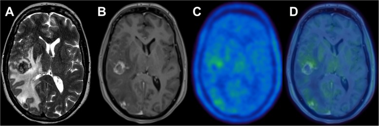
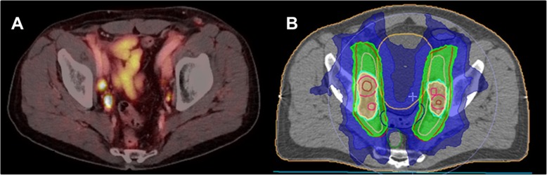
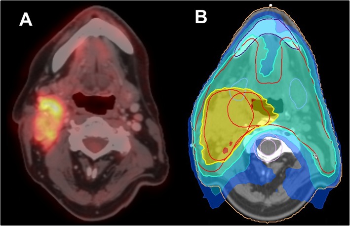
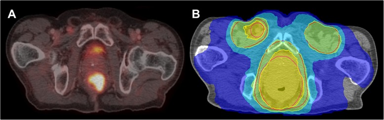
Radiotherapy and radiation oncology play a key role in the clinical management of patients suffering from oncological diseases. In clinical routine, anatomic imaging such as contrast-enhanced CT and MRI are widely available and are usually used to improve the target volume delineation for subsequent radiotherapy. Moreover, these modalities are also used for treatment monitoring after radiotherapy. However, some diagnostic questions cannot be sufficiently addressed by the mere use standard morphological imaging. Therefore, positron emission tomography (PET) imaging gains increasing clinical significance in the management of oncological patients undergoing radiotherapy, as PET allows the visualization and quantification of tumoral features on a molecular level beyond the mere morphological extent shown by conventional imaging, such as tumor metabolism or receptor expression. The tumor metabolism or receptor expression information derived from PET can be used as tool for visualization of tumor extent, for assessing response during and after therapy, for prediction of patterns of failure and for definition of the volume in need of dose-escalation. This review focuses on recent and current advances of PET imaging within the field of clinical radiotherapy / radiation oncology in several oncological entities (neuro-oncology, head & neck cancer, lung cancer, gastrointestinal tumors and prostate cancer) with particular emphasis on radiotherapy planning, response assessment after radiotherapy and prognostication.
放射治疗和放射肿瘤学在治疗肿瘤疾病患者的临床管理中起着关键作用。在临床常规中,解剖成像(如增强 CT 和 MRI)广泛可用,通常用于改善随后放射治疗的靶区勾画。此外,这些方式也用于放射治疗后的治疗监测。然而,仅使用标准形态成像并不能充分解决某些诊断问题。因此,正电子发射断层扫描(PET)成像在接受放射治疗的肿瘤患者的管理中获得了越来越重要的临床意义,因为 PET 允许在分子水平上可视化和量化肿瘤特征,而不仅仅是常规成像显示的形态学范围,如肿瘤代谢或受体表达。从 PET 获得的肿瘤代谢或受体表达信息可用于可视化肿瘤范围、评估治疗期间和治疗后的反应、预测失败模式以及定义需要剂量递增的体积。这篇综述重点介绍了在神经肿瘤学、头颈部癌症、肺癌、胃肠道肿瘤和前列腺癌等几种肿瘤实体中,临床放射治疗/放射肿瘤学领域中 PET 成像的最新和当前进展,特别强调了放射治疗计划、放射治疗后反应评估和预后。