Caraiani Cosmin, Yi Dong, Petresc Bianca, Dietrich Christoph
Department of Medical Imaging, "Iuliu Hatieganu" University of Medicine and Pharmacy , Cluj-Napoca , Romania.
Department of Ultrasound, Zhongshan Hospital, Fudan University , Shanghai , China.
J Ultrason. 2020;20(80):e43-e54. doi: 10.15557/JoU.2020.0008. Epub 2020 Mar 31.
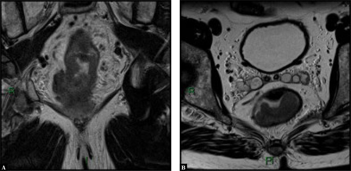
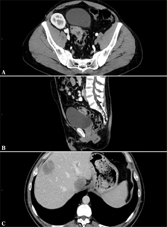
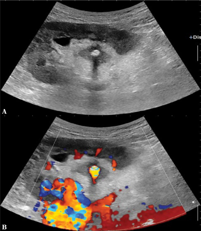
Imaging has a very important role in evaluating abdominal pathology. A good knowledge of indications is of crucial importance in the management of the patient with abdominal pathology. Ultrasound, which on its own can lead to an accurate diagnosis, plays a pivotal role in the management of abdominal pathology. The use of ultrasound contrast agents has significantly improved ultrasound diagnostic capacities in both hepatic and non-hepatic pathology. The use of computed tomography should be limited due to the potential harmful side effects of ionizing radiation, but it has established roles in evaluating severe abdominal traumatic and non-traumatic emergencies as well as in staging oncologic patients. Magnetic resonance imaging has very limited utility in abdominal emergencies due to difficulty of accessing the scanner and the long duration of the examination compared to computed tomography or ultrasound. However, magnetic resonance imaging has well-established clinical roles particularly for evaluating diffuse or focal hepatic pathology, benign and malignant bile duct pathology, pancreatic tumors, inflammatory bowel disease and rectal tumors. The aims of the following paper are to familiarize the clinician with the indications for imaging in abdominal pathology, to guide the clinician and radiologist in choosing the correct technique for a particular clinical situation, to prevent the overuse of imaging techniques and to prevent misdiagnosis of disease and incorrect therapy resulting from inappropriate imaging. Imaging has a very important role in evaluating abdominal pathology. A good knowledge of indications is of crucial importance in the management of the patient with abdominal pathology. Ultrasound, which on its own can lead to an accurate diagnosis, plays a pivotal role in the management of abdominal pathology. The use of ultrasound contrast agents has significantly improved ultrasound diagnostic capacities in both hepatic and non-hepatic pathology. The use of computed tomography should be limited due to the potential harmful side effects of ionizing radiation, but it has established roles in evaluating severe abdominal traumatic and non-traumatic emergencies as well as in staging oncologic patients. Magnetic resonance imaging has very limited utility in abdominal emergencies due to difficulty of accessing the scanner and the long duration of the examination compared to computed tomography or ultrasound. However, magnetic resonance imaging has well-established clinical roles particularly for evaluating diffuse or focal hepatic pathology, benign and malignant bile duct pathology, pancreatic tumors, inflammatory bowel disease and rectal tumors. The aims of the following paper are to familiarize the clinician with the indications for imaging in abdominal pathology, to guide the clinician and radiologist in choosing the correct technique for a particular clinical situation, to prevent the overuse of imaging techniques and to prevent misdiagnosis of disease and incorrect therapy resulting from inappropriate imaging.
影像学在评估腹部病变中起着非常重要的作用。熟悉适应证对于腹部病变患者的管理至关重要。超声本身就能得出准确诊断,在腹部病变的管理中发挥着关键作用。超声造影剂的使用显著提高了超声在肝脏和非肝脏病变中的诊断能力。由于电离辐射潜在的有害副作用,计算机断层扫描(CT)的使用应受到限制,但它在评估严重腹部创伤和非创伤性急症以及肿瘤患者分期方面具有既定作用。由于与CT或超声相比,磁共振成像(MRI)存在难以进入检查设备以及检查时间长的问题,其在腹部急症中的应用非常有限。然而,MRI在评估弥漫性或局灶性肝脏病变、良性和恶性胆管病变、胰腺肿瘤、炎症性肠病和直肠肿瘤方面具有明确的临床作用。本文的目的是使临床医生熟悉腹部病变的影像学适应证,指导临床医生和放射科医生针对特定临床情况选择正确技术,防止影像学技术的过度使用,并防止因不恰当的影像学检查导致疾病误诊和治疗错误。影像学在评估腹部病变中起着非常重要的作用。熟悉适应证对于腹部病变患者的管理至关重要。超声本身就能得出准确诊断,在腹部病变的管理中发挥着关键作用。超声造影剂的使用显著提高了超声在肝脏和非肝脏病变中的诊断能力。由于电离辐射潜在的有害副作用,计算机断层扫描(CT)的使用应受到限制,但它在评估严重腹部创伤和非创伤性急症以及肿瘤患者分期方面具有既定作用。由于与CT或超声相比,磁共振成像(MRI)存在难以进入检查设备以及检查时间长的问题,其在腹部急症中的应用非常有限。然而,MRI在评估弥漫性或局灶性肝脏病变、良性和恶性胆管病变、胰腺肿瘤、炎症性肠病和直肠肿瘤方面具有明确的临床作用。本文的目的是使临床医生熟悉腹部病变的影像学适应证,指导临床医生和放射科医生针对特定临床情况选择正确技术,防止影像学技术的过度使用,并防止因不恰当的影像学检查导致疾病误诊和治疗错误。