Department of Theory and Bio-Systems, Max Planck Institute of Colloids and Interfaces, Science Park Golm, 14424 Potsdam, Germany.
Nano Lett. 2020 May 13;20(5):3185-3191. doi: 10.1021/acs.nanolett.9b05232. Epub 2020 Apr 29.
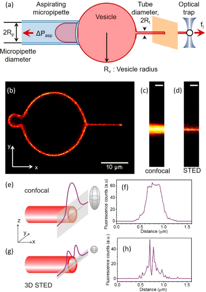
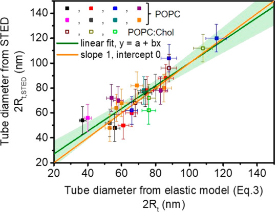
Membrane tension modulates the morphology of plasma-membrane tubular protrusions in cells but is difficult to measure. Here, we propose to use microscopy imaging to assess the membrane tension. We report direct measurement of membrane nanotube diameters with unprecedented resolution using stimulated emission depletion (STED) microscopy. For this purpose, we integrated an optical tweezers setup in a commercial microscope equipped for STED imaging and established micropipette aspiration of giant vesicles. Membrane nanotubes were pulled from the vesicles at specific membrane tension imposed by the aspiration pipet. Tube diameters calculated from the applied tension using the membrane curvature elasticity model are in excellent agreement with data measured directly with STED. Our approach can be extended to cellular membranes and will then allow us to estimate the mechanical membrane tension within the force-induced nanotubes.
膜张力调节细胞中质膜管状突起的形态,但很难测量。在这里,我们建议使用显微镜成像来评估膜张力。我们报告了使用受激发射损耗(STED)显微镜以空前的分辨率直接测量膜纳米管直径。为此,我们将光学镊子装置集成到配备 STED 成像的商用显微镜中,并建立了巨囊泡的微量吸管抽吸。通过抽吸微管在特定的膜张力下从囊泡中拉出膜纳米管。根据膜曲率弹性模型从所施加的张力计算得到的管直径与使用 STED 直接测量的数据非常吻合。我们的方法可以扩展到细胞膜,并允许我们估计力诱导纳米管内的机械膜张力。