• 文献检索
  • 文档翻译
  • 深度研究
  • 学术资讯
  • Suppr Zotero 插件Zotero 插件
  • 邀请有礼
  • 套餐&价格
  • 历史记录
应用&插件
Suppr Zotero 插件Zotero 插件浏览器插件Mac 客户端Windows 客户端微信小程序
定价
高级版会员购买积分包购买API积分包
服务
文献检索文档翻译深度研究API 文档MCP 服务
关于我们
关于 Suppr公司介绍联系我们用户协议隐私条款
关注我们

Suppr 超能文献

核心技术专利:CN118964589B侵权必究
粤ICP备2023148730 号-1Suppr @ 2026

文献检索

告别复杂PubMed语法,用中文像聊天一样搜索,搜遍4000万医学文献。AI智能推荐,让科研检索更轻松。

立即免费搜索

文件翻译

保留排版,准确专业,支持PDF/Word/PPT等文件格式,支持 12+语言互译。

免费翻译文档

深度研究

AI帮你快速写综述,25分钟生成高质量综述,智能提取关键信息,辅助科研写作。

立即免费体验

基于时空分布的致痫性脑源的无创电磁场源成像

Noninvasive electromagnetic source imaging of spatiotemporally distributed epileptogenic brain sources.

机构信息

Department of Biomedical Engineering, Carnegie Mellon University, Pittsburgh, PA, USA.

Department of Biomedical Engineering, University of Minnesota, Minnesota, MN, USA.

出版信息

Nat Commun. 2020 Apr 23;11(1):1946. doi: 10.1038/s41467-020-15781-0.

DOI:10.1038/s41467-020-15781-0
PMID:32327635
原文链接:https://pmc.ncbi.nlm.nih.gov/articles/PMC7181775/
Abstract

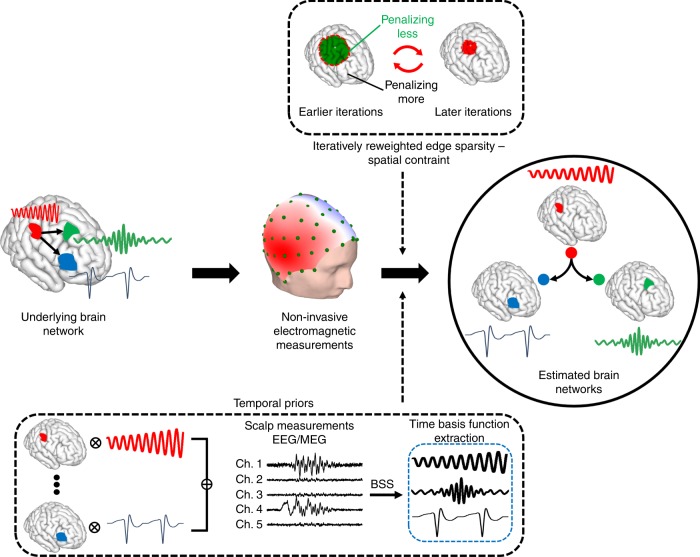
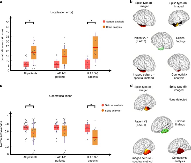
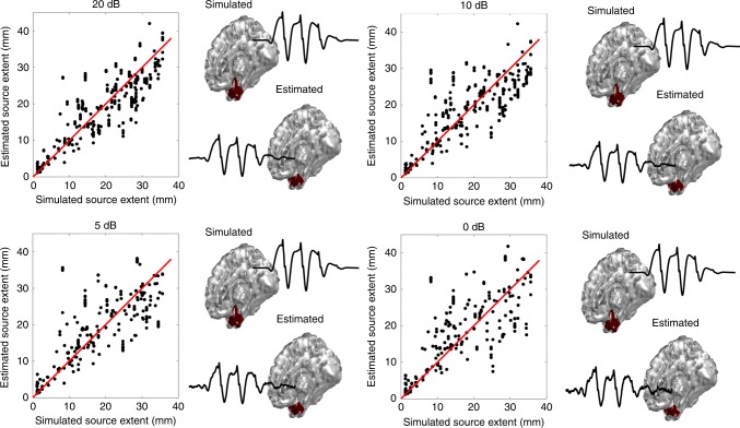
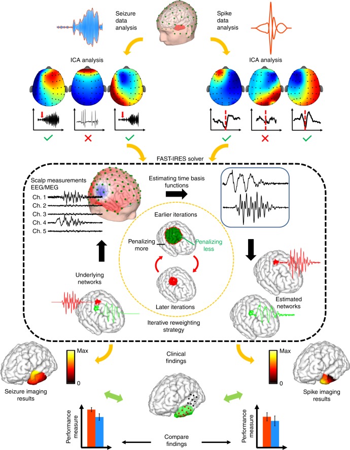
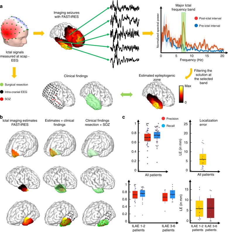
Brain networks are spatiotemporal phenomena that dynamically vary over time. Functional imaging approaches strive to noninvasively estimate these underlying processes. Here, we propose a novel source imaging approach that uses high-density EEG recordings to map brain networks. This approach objectively addresses the long-standing limitations of conventional source imaging techniques, namely, difficulty in objectively estimating the spatial extent, as well as the temporal evolution of underlying brain sources. We validate our approach by directly comparing source imaging results with the intracranial EEG (iEEG) findings and surgical resection outcomes in a cohort of 36 patients with focal epilepsy. To this end, we analyzed a total of 1,027 spikes and 86 seizures. We demonstrate the capability of our approach in imaging both the location and spatial extent of brain networks from noninvasive electrophysiological measurements, specifically for ictal and interictal brain networks. Our approach is a powerful tool for noninvasively investigating large-scale dynamic brain networks.

摘要

脑网络是时空现象,随时间动态变化。功能成像方法旨在无创地估计这些潜在过程。在这里,我们提出了一种新的源成像方法,使用高密度 EEG 记录来绘制脑网络。该方法客观地解决了传统源成像技术的长期局限性,即难以客观估计潜在脑源的空间范围和时间演变。我们通过在 36 例局灶性癫痫患者的队列中直接将源成像结果与颅内 EEG(iEEG)发现和手术切除结果进行比较来验证我们的方法。为此,我们分析了总共 1027 个尖峰和 86 个发作。我们展示了我们的方法在从非侵入性电生理测量中成像脑网络的位置和空间范围的能力,特别是对于发作期和发作间期脑网络。我们的方法是一种用于无创研究大规模动态脑网络的强大工具。

https://cdn.ncbi.nlm.nih.gov/pmc/blobs/f3c2/7181775/da9f361cb9a8/41467_2020_15781_Fig6_HTML.jpg
https://cdn.ncbi.nlm.nih.gov/pmc/blobs/f3c2/7181775/846b01cb63cc/41467_2020_15781_Fig1_HTML.jpg
https://cdn.ncbi.nlm.nih.gov/pmc/blobs/f3c2/7181775/8ebbc9afe46a/41467_2020_15781_Fig2_HTML.jpg
https://cdn.ncbi.nlm.nih.gov/pmc/blobs/f3c2/7181775/b858afc2881d/41467_2020_15781_Fig3_HTML.jpg
https://cdn.ncbi.nlm.nih.gov/pmc/blobs/f3c2/7181775/40141b8e8acc/41467_2020_15781_Fig4_HTML.jpg
https://cdn.ncbi.nlm.nih.gov/pmc/blobs/f3c2/7181775/124949ca5f68/41467_2020_15781_Fig5_HTML.jpg
https://cdn.ncbi.nlm.nih.gov/pmc/blobs/f3c2/7181775/da9f361cb9a8/41467_2020_15781_Fig6_HTML.jpg

相似文献

1
Noninvasive electromagnetic source imaging of spatiotemporally distributed epileptogenic brain sources.基于时空分布的致痫性脑源的无创电磁场源成像
Nat Commun. 2020 Apr 23;11(1):1946. doi: 10.1038/s41467-020-15781-0.
2
Electric source imaging of interictal activity accurately localises the seizure onset zone.电源成像技术能够准确地定位发作间期活动的起源区。
J Neurol Neurosurg Psychiatry. 2014 Jan;85(1):38-43. doi: 10.1136/jnnp-2013-305515. Epub 2013 Jul 30.
3
Localization Accuracy of Distributed Inverse Solutions for Electric and Magnetic Source Imaging of Interictal Epileptic Discharges in Patients with Focal Epilepsy.局灶性癫痫患者发作间期癫痫放电的电和磁源成像分布式逆解的定位准确性
Brain Topogr. 2016 Jan;29(1):162-81. doi: 10.1007/s10548-014-0423-1. Epub 2015 Jan 22.
4
Identifying epileptic source location and extent: an iterative sparse electromagnetic source imaging algorithm.识别癫痫源的位置和范围:一种迭代稀疏电磁源成像算法。
Annu Int Conf IEEE Eng Med Biol Soc. 2016 Aug;2016:109-112. doi: 10.1109/EMBC.2016.7590652.
5
Noninvasive imaging of the high frequency brain activity in focal epilepsy patients.局灶性癫痫患者高频脑活动的无创成像
IEEE Trans Biomed Eng. 2014 Jun;61(6):1660-7. doi: 10.1109/TBME.2013.2297332.
6
Interictal and ictal source localization for epilepsy surgery using high-density EEG with MEG: a prospective long-term study.应用高密度脑电图和脑磁图进行癫痫手术的发作间期和发作期源定位:一项前瞻性长期研究。
Brain. 2019 Apr 1;142(4):932-951. doi: 10.1093/brain/awz015.
7
Evaluation of algorithms for intracranial EEG (iEEG) source imaging of extended sources: feasibility of using iEEG source imaging for localizing epileptogenic zones in secondary generalized epilepsy.评估扩展源颅内 EEG(iEEG)源成像算法:使用 iEEG 源成像定位继发性全面性癫痫发作区的可行性。
Brain Topogr. 2011 Jun;24(2):91-104. doi: 10.1007/s10548-011-0173-2. Epub 2011 Mar 3.
8
Imaging the extent and location of spatiotemporally distributed epileptiform sources from MEG measurements.从脑磁图测量中对时空分布的癫痫源的范围和位置进行成像。
Neuroimage Clin. 2022;33:102903. doi: 10.1016/j.nicl.2021.102903. Epub 2021 Nov 29.
9
Defining epileptogenic networks: Contribution of SEEG and signal analysis.定义致痫网络:SEEG 和信号分析的贡献。
Epilepsia. 2017 Jul;58(7):1131-1147. doi: 10.1111/epi.13791. Epub 2017 May 20.
10
Preoperative simulation of intracerebral epileptiform discharges: synthetic aperture magnetometry virtual sensor analysis of interictal magnetoencephalography data.脑内癫痫样放电的术前模拟:发作间期脑磁图数据的合成孔径磁力测定虚拟传感器分析
J Neurosurg. 2006 Jul;105(1 Suppl):41-9. doi: 10.3171/ped.2006.105.1.41.

引用本文的文献

1
Dynamic Mapping of Ictal Source Propagation From Stereo-EEG.基于立体脑电图的发作期源传播动态映射
Epilepsy Curr. 2025 Aug 3:15357597251365739. doi: 10.1177/15357597251365739.
2
Post-processing of a distributed source method for the localization of somatosensory cortex in a cohort of epilepsy patients.癫痫患者队列中体感皮层定位的分布式源方法的后处理
Neuroimage Rep. 2024 May 9;4(2):100204. doi: 10.1016/j.ynirp.2024.100204. eCollection 2024 Jun.
3
Stereo-EEG propagating source reconstruction identifies new surgical targets for epilepsy patients.

本文引用的文献

1
Deep brain activities can be detected with magnetoencephalography.深部脑活动可以用脑磁图进行检测。
Nat Commun. 2019 Feb 27;10(1):971. doi: 10.1038/s41467-019-08665-5.
2
Subcortical electrophysiological activity is detectable with high-density EEG source imaging.皮层下电生理活动可以通过高密度 EEG 源成像检测到。
Nat Commun. 2019 Feb 14;10(1):753. doi: 10.1038/s41467-019-08725-w.
3
Electrophysiological Source Imaging: A Noninvasive Window to Brain Dynamics.电生理源成像:窥探大脑动态的无创之窗。
立体脑电图传播源重建为癫痫患者确定了新的手术靶点。
Brain. 2025 Mar 6;148(3):764-775. doi: 10.1093/brain/awae297.
4
Flexible Alternating Projection for Spatially Extended Brain Source Localization.用于空间扩展脑源定位的灵活交替投影法
IEEE Trans Biomed Eng. 2025 Apr;72(4):1486-1497. doi: 10.1109/TBME.2024.3509741. Epub 2025 Mar 21.
5
Neuronavigation-Guided Transcranial Acoustoelectric Brain Imaging: A New Modality for High Resolution Electrical Brain Mapping.神经导航引导的经颅声电脑成像:一种用于高分辨率脑电图谱绘制的新方法。
IEEE Trans Biomed Eng. 2025 May;72(5):1605-1614. doi: 10.1109/TBME.2024.3514553. Epub 2025 Apr 23.
6
Low-density scalp electrical source imaging of the ictal onset zone network using source coherence maps.使用源相干图对发作起始区网络进行低密度头皮电源成像。
Front Neurol. 2024 Dec 19;15:1483977. doi: 10.3389/fneur.2024.1483977. eCollection 2024.
7
Robust interpolation of EEG/MEG sensor time-series via electromagnetic source imaging.通过电磁源成像对脑电图/脑磁图传感器时间序列进行稳健插值
J Neural Eng. 2025 Jan 15;22(1). doi: 10.1088/1741-2552/ada309.
8
Cortex level connectivity between ACT-R modules during EEG-based n-back task.基于脑电图的n-back任务期间ACT-R模块之间的皮质水平连通性。
Cogn Neurodyn. 2024 Dec;18(6):4033-4045. doi: 10.1007/s11571-024-10177-y. Epub 2024 Oct 21.
9
M/EEG source localization for both subcortical and cortical sources using a convolutional neural network with a realistic head conductivity model.使用具有逼真头部电导率模型的卷积神经网络对皮层下和皮层源进行脑磁图/脑电图源定位。
APL Bioeng. 2024 Oct 28;8(4):046104. doi: 10.1063/5.0226457. eCollection 2024 Dec.
10
Seizure Sources Can Be Imaged from Scalp EEG by Means of Biophysically Constrained Deep Neural Networks.癫痫发作源可通过生物物理约束深度神经网络从头皮脑电图成像。
Adv Sci (Weinh). 2024 Dec;11(47):e2405246. doi: 10.1002/advs.202405246. Epub 2024 Oct 29.
Annu Rev Biomed Eng. 2018 Jun 4;20:171-196. doi: 10.1146/annurev-bioeng-062117-120853. Epub 2018 Mar 1.
4
SISSY: An efficient and automatic algorithm for the analysis of EEG sources based on structured sparsity.SISSY:一种基于结构稀疏性的 EEG 源分析的高效自动算法。
Neuroimage. 2017 Aug 15;157:157-172. doi: 10.1016/j.neuroimage.2017.05.046. Epub 2017 May 31.
5
Individualised prediction model of seizure recurrence and long-term outcomes after withdrawal of antiepileptic drugs in seizure-free patients: a systematic review and individual participant data meta-analysis.在无癫痫发作的患者中停用抗癫痫药物后癫痫复发和长期结局的个体化预测模型:系统评价和个体参与者数据荟萃分析。
Lancet Neurol. 2017 Jul;16(7):523-531. doi: 10.1016/S1474-4422(17)30114-X. Epub 2017 May 5.
6
Source localization of ictal epileptic activity based on high-density scalp EEG data.基于高密度头皮 EEG 数据的发作期癫痫活动的源定位。
Epilepsia. 2017 Jun;58(6):1027-1036. doi: 10.1111/epi.13749. Epub 2017 Apr 11.
7
Diagnostic utility of invasive EEG for epilepsy surgery: Indications, modalities, and techniques.侵入性脑电图在癫痫手术中的诊断效用:适应症、方式和技术。
Epilepsia. 2016 Nov;57(11):1735-1747. doi: 10.1111/epi.13515. Epub 2016 Sep 28.
8
Complex patterns of spatially extended generators of epileptic activity: Comparison of source localization methods cMEM and 4-ExSo-MUSIC on high resolution EEG and MEG data.复杂的空间扩展癫痫活动发生器模式:高分辨率 EEG 和 MEG 数据上 cMEM 和 4-ExSo-MUSIC 源定位方法的比较。
Neuroimage. 2016 Dec;143:175-195. doi: 10.1016/j.neuroimage.2016.08.044. Epub 2016 Aug 22.
9
Imaging brain source extent from EEG/MEG by means of an iteratively reweighted edge sparsity minimization (IRES) strategy.通过迭代加权边缘稀疏性最小化(IRES)策略从脑电图/脑磁图成像脑源范围。
Neuroimage. 2016 Nov 15;142:27-42. doi: 10.1016/j.neuroimage.2016.05.064. Epub 2016 May 27.
10
Brain imaging in the assessment for epilepsy surgery.癫痫手术评估中的脑成像
Lancet Neurol. 2016 Apr;15(4):420-33. doi: 10.1016/S1474-4422(15)00383-X. Epub 2016 Feb 24.