Signal Processing and Recognition Group, Universidad Nacional de Colombia, Manizales, Colombia.
Instituto Tecnológico Metropolitano, Medellín, Colombia.
Comput Math Methods Med. 2020 Apr 3;2020:5076865. doi: 10.1155/2020/5076865. eCollection 2020.
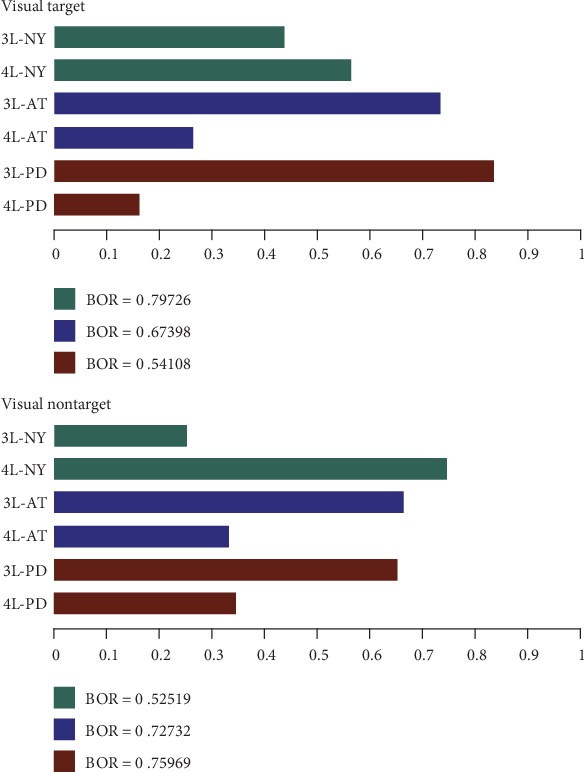
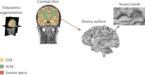
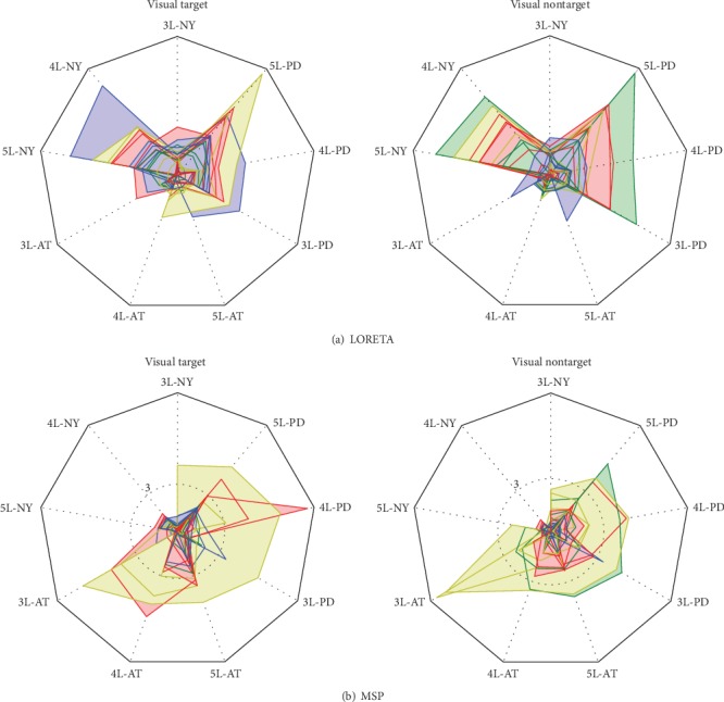
Electromagnetic source imaging (ESI) techniques have become one of the most common alternatives for understanding cognitive processes in the human brain and for guiding possible therapies for neurological diseases. However, ESI accuracy strongly depends on the forward model capabilities to accurately describe the subject's head anatomy from the available structural data. Attempting to improve the ESI performance, we enhance the brain structure model within the individual-defined forward problem formulation, combining the head geometry complexity of the modeled tissue compartments and the prior knowledge of the brain tissue morphology. We validate the proposed methodology using 25 subjects, from which a set of magnetic-resonance imaging scans is acquired, extracting the anatomical priors and an electroencephalography signal set needed for validating the ESI scenarios. Obtained results confirm that incorporating patient-specific head models enhances the performed accuracy and improves the localization of focal and deep sources.
电磁源成像(ESI)技术已成为了解人类大脑认知过程和指导神经疾病可能治疗方法的最常用方法之一。然而,ESI 的准确性在很大程度上取决于正向模型的能力,即能否从可用的结构数据中准确描述受试者的头部解剖结构。为了提高 ESI 的性能,我们在个体定义的正向问题公式中增强了大脑结构模型,将建模组织隔室的头部几何复杂性与大脑组织形态的先验知识相结合。我们使用 25 名受试者验证了所提出的方法,从这些受试者中获取了一组磁共振成像扫描,提取了进行 ESI 场景验证所需的解剖先验和脑电图信号集。得到的结果证实,纳入患者特定的头部模型可以提高准确性,并改善焦点和深部源的定位。