Department of Biomedical Engineering, The University of Texas, Austin, TX, USA.
LiveSTRONG Cancer Institutes, The University of Texas, Austin, TX, USA.
BMC Cancer. 2020 Apr 28;20(1):359. doi: 10.1186/s12885-020-06868-4.
Therapy targeted to the human epidermal growth factor receptor type 2 (HER2) is used in combination with cytotoxic therapy in treatment of HER2+ breast cancer. Trastuzumab, a monoclonal antibody that targets HER2, has been shown pre-clinically to induce vascular changes that can increase delivery of chemotherapy. To quantify the role of immune modulation in treatment-induced vascular changes, this study identifies temporal changes in myeloid cell infiltration with corresponding vascular alterations in a preclinical model of HER2+ breast cancer following trastuzumab treatment.
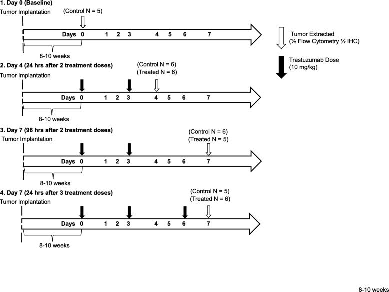
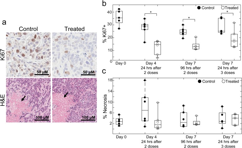
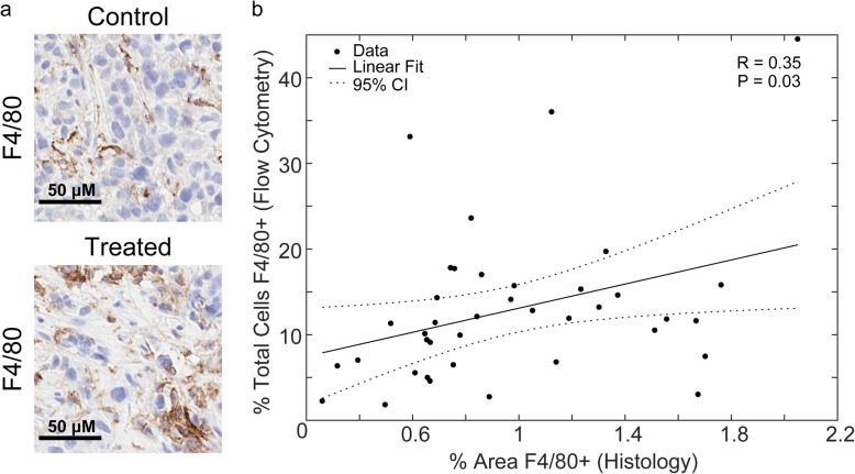
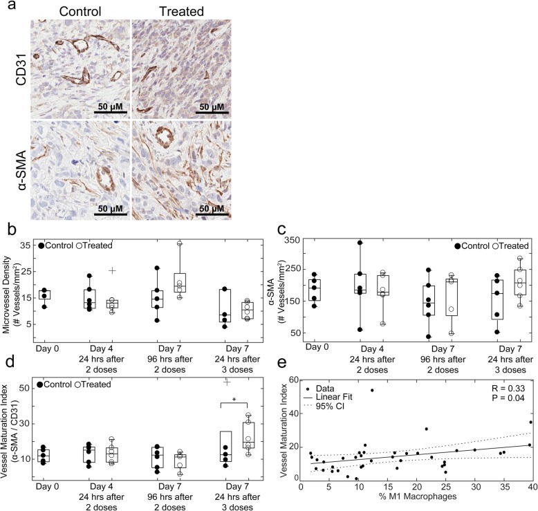
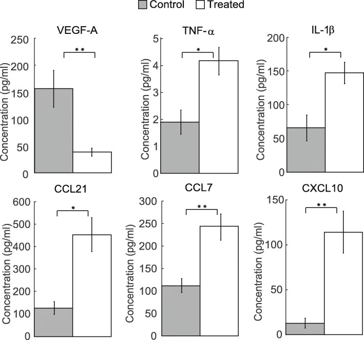
HER2+ tumor-bearing mice (N = 46) were treated with trastuzumab or saline. After extraction, half of each tumor was analyzed by immunophenotyping using flow cytometry. The other half was quantified by immunohistochemistry to characterize macrophage infiltration (F4/80), vascularity (CD31 and α-SMA), proliferation (Ki67) and cellularity (H&E). Additional mice (N = 10) were used to quantify differences in tumor cytokines between control and treated groups.
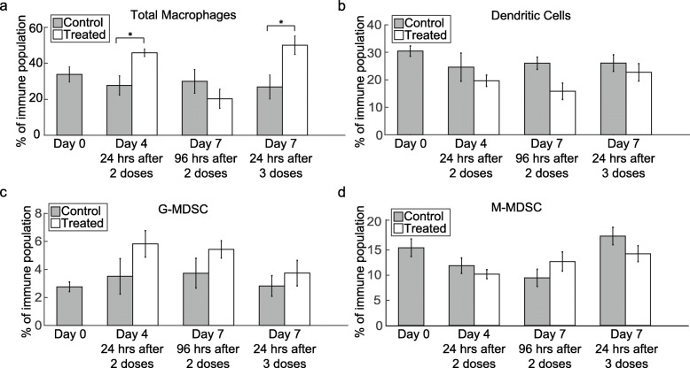
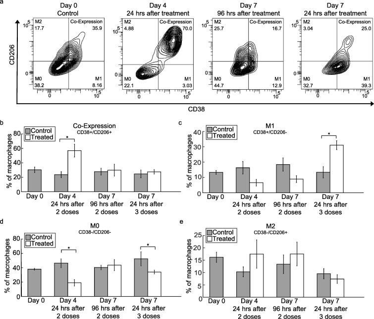
Immunophenotyping showed an increase in macrophage infiltration 24 h after trastuzumab treatment (P ≤ 0.05). With continued trastuzumab treatment, the M1 macrophage population increased (P = 0.02). Increases in vessel maturation index (i.e., the ratio of α-SMA to CD31) positively correlated with increases in tumor infiltrating M1 macrophages (R = 0.33, P = 0.04). Decreases in VEGF-A and increases in inflammatory cytokines (TNF-α, IL-1β, CCL21, CCL7, and CXCL10) were observed with continued trastuzumab treatment (P ≤ 0.05).
Preliminary results from this study in a murine model of HER2+ breast cancer show correlations between immune modulation and vascular changes, and reveals the potential for anti-HER2 therapy to reprogram immunosuppressive components of the tumor microenvironment. The quantification of immune modulation in HER2+ breast cancer, as well as the mechanistic insight of vascular alterations after anti-HER2 treatment, represent novel contributions and warrant further assessment for potential clinical translation.
针对人表皮生长因子受体 2(HER2)的治疗方法与细胞毒性疗法联合用于治疗 HER2+乳腺癌。曲妥珠单抗是一种针对 HER2 的单克隆抗体,已在临床前研究中显示可诱导血管变化,从而增加化疗药物的递送。为了量化免疫调节在治疗诱导的血管变化中的作用,本研究在曲妥珠单抗治疗后,通过识别 HER2+乳腺癌的临床前模型中髓样细胞浸润的时间变化,以及相应的血管改变,来确定免疫调节的作用。
HER2+荷瘤小鼠(N=46)接受曲妥珠单抗或生理盐水治疗。提取后,一半肿瘤通过流式细胞术免疫表型分析进行分析。另一半通过免疫组织化学进行定量分析,以表征巨噬细胞浸润(F4/80)、血管生成(CD31 和 α-SMA)、增殖(Ki67)和细胞密度(H&E)。另外 10 只小鼠(N=10)用于定量分析对照组和治疗组之间肿瘤细胞因子的差异。
免疫表型分析显示,曲妥珠单抗治疗后 24 小时巨噬细胞浸润增加(P≤0.05)。随着曲妥珠单抗治疗的继续,M1 巨噬细胞群增加(P=0.02)。血管成熟指数(即 α-SMA 与 CD31 的比值)的增加与肿瘤浸润 M1 巨噬细胞的增加呈正相关(R=0.33,P=0.04)。随着曲妥珠单抗治疗的继续,VEGF-A 减少,炎症细胞因子(TNF-α、IL-1β、CCL21、CCL7 和 CXCL10)增加(P≤0.05)。
本研究在 HER2+乳腺癌的小鼠模型中初步结果表明,免疫调节与血管变化之间存在相关性,并揭示了抗 HER2 治疗重新编程肿瘤微环境中免疫抑制成分的潜力。HER2+乳腺癌中免疫调节的定量以及抗 HER2 治疗后血管改变的机制见解是新的贡献,值得进一步评估以潜在转化为临床应用。