Clinical Pathology Unit and Center of Molecular Medicine, Department of Medical and Surgical Sciences, University of Foggia, Viale Luigi Pinto 71122, Foggia, Italy.
Urology and Renal Transplantation Unit, Department of Emergency and Organ Transplantation, University of Bari "Aldo Moro", Bari 70124, Italy.
Aging (Albany NY). 2020 Apr 28;12(8):7585-7602. doi: 10.18632/aging.103169.
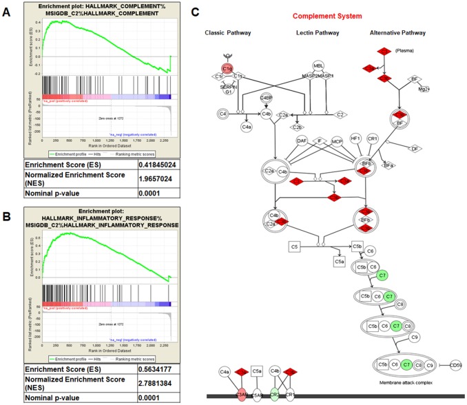
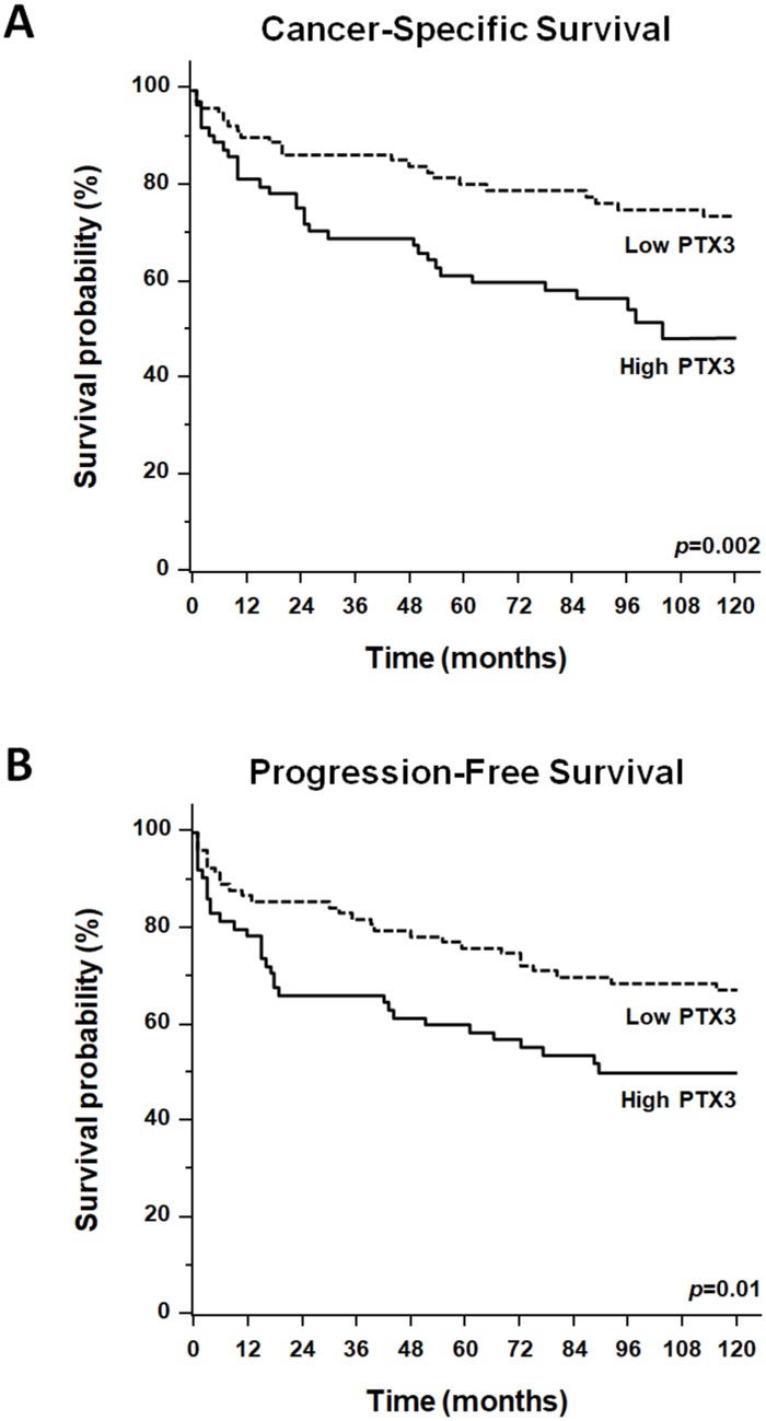
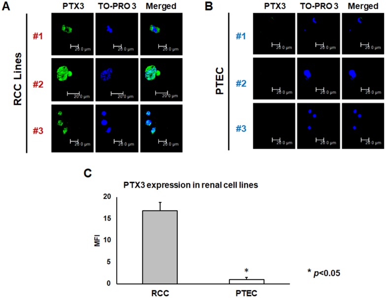
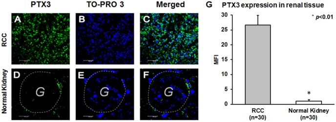
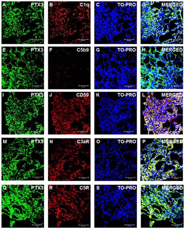
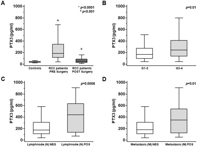
Pentraxin-3 (PTX3) belongs to the pentraxine family, innate immune regulators involved in angiogenesis, proliferation and immune escape in cancer. Here, we evaluated PTX3 tissue expression and serum levels as biomarkers of clear cell renal cell carcinoma (ccRCC) and analyzed the possible role of complement system activation on tumor site. A 10-year retrospective cohort study including patients undergoing nephrectomy for ccRCC was also performed. PTX3 expression was elevated in both neoplastic renal cell lines and tissues, while it was absent in both normal renal proximal tubular cells (HK2) and normal renal tissues. Analysis of complement system activation on tumor tissues showed the co-expression of PTX3 with C1q, C3aR, C5R1 and CD59, but not with C5b-9 terminal complex. RCC patients showed higher serum PTX3 levels as compared to non-neoplastic patients (p<0.0001). Higher PTX3 serum levels were observed in patients with higher Fuhrman grade (p<0.01), lymph node (p<0.0001), and visceral metastases (p<0.001). Patients with higher PTX3 levels also showed significantly lower survival rates (p=0.002). Our results suggest that expression of PTX3 can affect the immunoflogosis in the ccRCC microenvironment, by activating the classical pathway of CS (C1q) and releasing pro-angiogenic factors (C3a, C5a). The up-regulation of CD59 also inhibits the complement-mediated cellular lysis.
血清 pentraxin-3(PTX3)属于先天免疫调节 pentaxine 家族,参与肿瘤血管生成、增殖和免疫逃逸。在此,我们评估了 PTX3 组织表达和血清水平作为透明细胞肾细胞癌(ccRCC)的生物标志物,并分析了补体系统激活在肿瘤部位的可能作用。还进行了一项 10 年回顾性队列研究,包括接受肾切除术治疗 ccRCC 的患者。PTX3 在肿瘤肾细胞系和组织中表达上调,而在正常肾近端管状细胞(HK2)和正常肾组织中均不存在。对肿瘤组织中补体系统激活的分析表明,PTX3 与 C1q、C3aR、C5R1 和 CD59 共表达,但不与 C5b-9 末端复合物共表达。与非肿瘤患者相比,RCC 患者的血清 PTX3 水平更高(p<0.0001)。PTX3 血清水平较高的患者 Fuhrman 分级较高(p<0.01)、淋巴结转移(p<0.0001)和内脏转移(p<0.001)。PTX3 水平较高的患者生存率也显著降低(p=0.002)。我们的结果表明,PTX3 的表达可以通过激活补体经典途径(C1q)和释放促血管生成因子(C3a、C5a)来影响 ccRCC 微环境中的免疫球蛋白,上调 CD59 还抑制补体介导的细胞溶解。