Department of Gastrointestinal Surgery, The Affiliated Yantai Yuhuangding Hospital of Qingdao University, Yantai, Shandong, China (mainland).
Med Sci Monit. 2020 May 1;26:e921383. doi: 10.12659/MSM.921383.
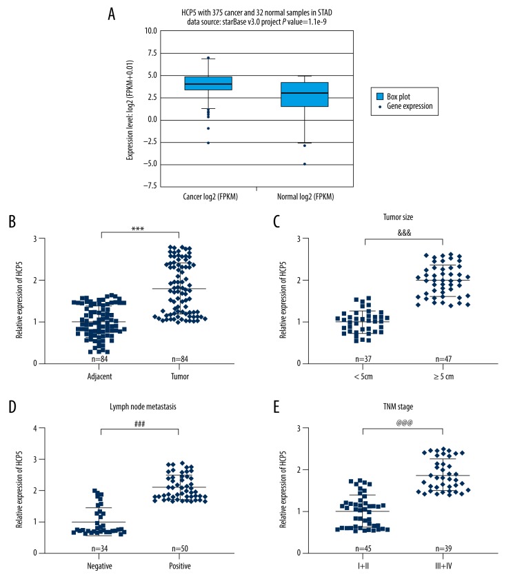
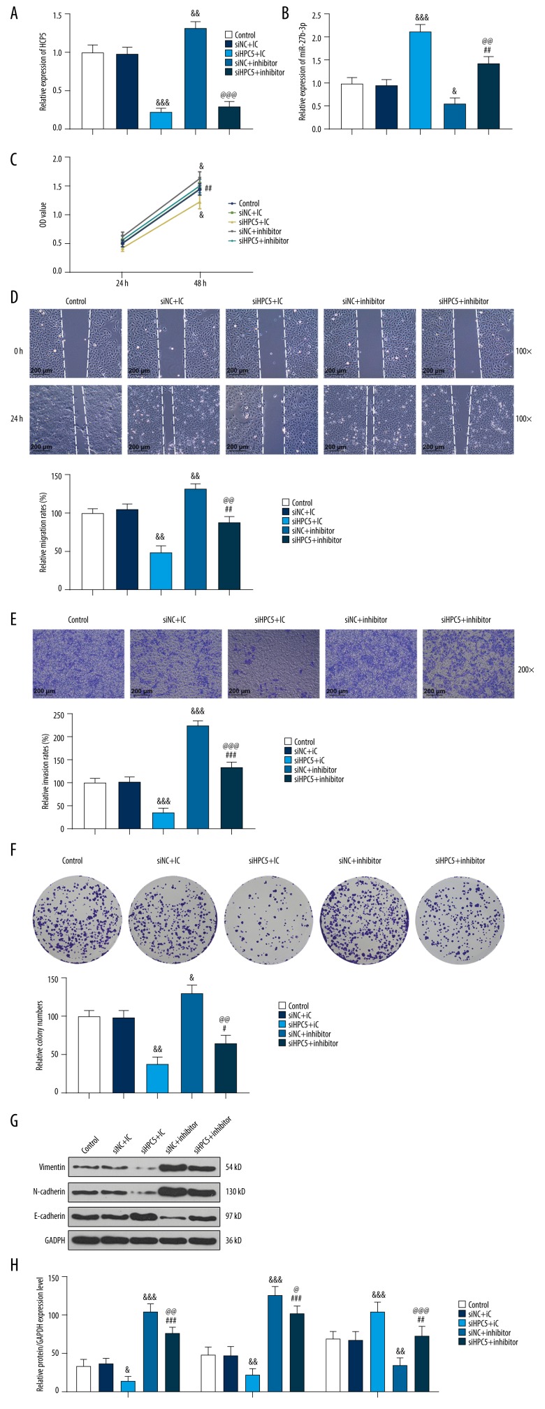
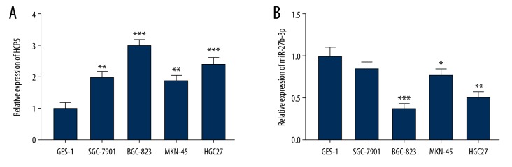
BACKGROUND lncRNA HCP5 plays a cancer-promoting role in a variety of cancers. This study was the first to explore the mechanism of HCP5 in gastric carcinoma (GC). MATERIAL AND METHODS The differences in HCP5 between GC patients and healthy people were revealed in the TCGA database. The expression of HCP5 in GC tissues and adjacent tissues was compared by qRT-PCR. At the same time, the clinic pathological features of the patients were counted. Starbase and luciferase assay predicted and verified that miR-27b-3p is a targeted miRNA for HCP5. The expression of HCP5 and miR-27b-3p in various GC cells was detected by qRT-PCR. Cell viability and metastasis in different treatment groups were assessed by use of Cell Couting Kit-8 assay and clone formation assay, wound-healing assay, and transwell assay. Finally, expression of epithelial-mesenchymal transition (EMT)-associated markers was detected by Western blot. RESULTS We found that HCP5 was overexpressed in GC tissues. Patients with higher expression of HCP5 had larger tumors, were more likely to have lymph node metastasis, and had higher TNM stage. HCP5 was overexpressed in GC cells, but this was reversed by miR-27b-3p. Silencing HCP5 inhibited GC cell viability and metastasis by downregulating Vimentin and N-cadherin and up-regulating E-cadherin, but this effect was partially reversed by miR-27b-3p inhibitor. CONCLUSIONS The effect of silencing HCP5 on repressing GC cells viability and metastasis by regulating EMT-associated markers can be partially reversed by miR-27b-3p inhibitor.
lncRNA HCP5 在多种癌症中发挥致癌作用。本研究首次探讨了 HCP5 在胃癌(GC)中的作用机制。
在 TCGA 数据库中揭示了 GC 患者与健康人之间 HCP5 的差异。通过 qRT-PCR 比较 GC 组织和相邻组织中 HCP5 的表达。同时,统计了患者的临床病理特征。Starbase 和荧光素酶测定预测并验证了 miR-27b-3p 是 HCP5 的靶向 miRNA。通过 qRT-PCR 检测了各种 GC 细胞中 HCP5 和 miR-27b-3p 的表达。通过细胞计数试剂盒-8 检测、克隆形成检测、划痕愈合检测和 Transwell 检测评估不同处理组中细胞活力和转移情况。最后,通过 Western blot 检测上皮间质转化(EMT)相关标志物的表达。
我们发现 HCP5 在 GC 组织中表达上调。HCP5 表达较高的患者肿瘤较大,更有可能发生淋巴结转移,且 TNM 分期更高。GC 细胞中 HCP5 表达上调,但被 miR-27b-3p 逆转。沉默 HCP5 通过下调 Vimentin 和 N-cadherin 并上调 E-cadherin 抑制 GC 细胞活力和转移,但这种作用被 miR-27b-3p 抑制剂部分逆转。
沉默 HCP5 通过调节 EMT 相关标志物抑制 GC 细胞活力和转移的作用可以被 miR-27b-3p 抑制剂部分逆转。