• 文献检索
  • 文档翻译
  • 深度研究
  • 学术资讯
  • Suppr Zotero 插件Zotero 插件
  • 邀请有礼
  • 套餐&价格
  • 历史记录
应用&插件
Suppr Zotero 插件Zotero 插件浏览器插件Mac 客户端Windows 客户端微信小程序
定价
高级版会员购买积分包购买API积分包
服务
文献检索文档翻译深度研究API 文档MCP 服务
关于我们
关于 Suppr公司介绍联系我们用户协议隐私条款
关注我们

Suppr 超能文献

核心技术专利:CN118964589B侵权必究
粤ICP备2023148730 号-1Suppr @ 2026

文献检索

告别复杂PubMed语法,用中文像聊天一样搜索,搜遍4000万医学文献。AI智能推荐,让科研检索更轻松。

立即免费搜索

文件翻译

保留排版,准确专业,支持PDF/Word/PPT等文件格式,支持 12+语言互译。

免费翻译文档

深度研究

AI帮你快速写综述,25分钟生成高质量综述,智能提取关键信息,辅助科研写作。

立即免费体验

超声检查中四平面法在乳腺病变计算机辅助诊断中的评估:前瞻性单中心研究

Evaluation of the Quadri-Planes Method in Computer-Aided Diagnosis of Breast Lesions by Ultrasonography: Prospective Single-Center Study.

作者信息

Yongping Liang, Juan Zhang, Zhou Ping, Yongfeng Zhao, Liu Wengang, Shi Yifan

机构信息

The Xiangya Medical School, Central South University, Changsha, Hunan, China.

出版信息

JMIR Med Inform. 2020 May 5;8(5):e18251. doi: 10.2196/18251.

DOI:10.2196/18251
PMID:32369039
原文链接:https://pmc.ncbi.nlm.nih.gov/articles/PMC7238092/
Abstract

BACKGROUND

Computer-aided diagnosis (CAD) is a tool that can help radiologists diagnose breast lesions by ultrasonography. Previous studies have demonstrated that CAD can help reduce the incidence of missed diagnoses by radiologists. However, the optimal method to apply CAD to breast lesions using diagnostic planes has not been assessed.

OBJECTIVE

The aim of this study was to compare the performance of radiologists with different levels of experience when using CAD with the quadri-planes method to detect breast tumors.

METHODS

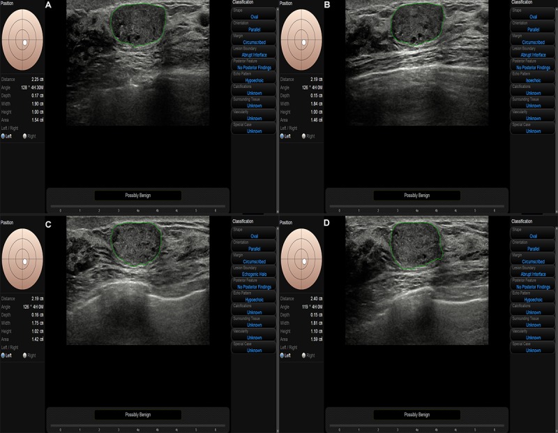
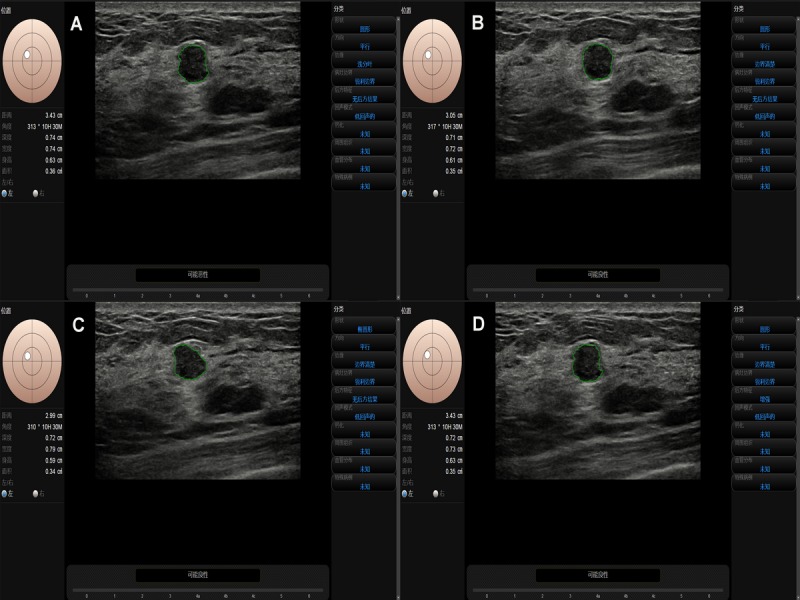
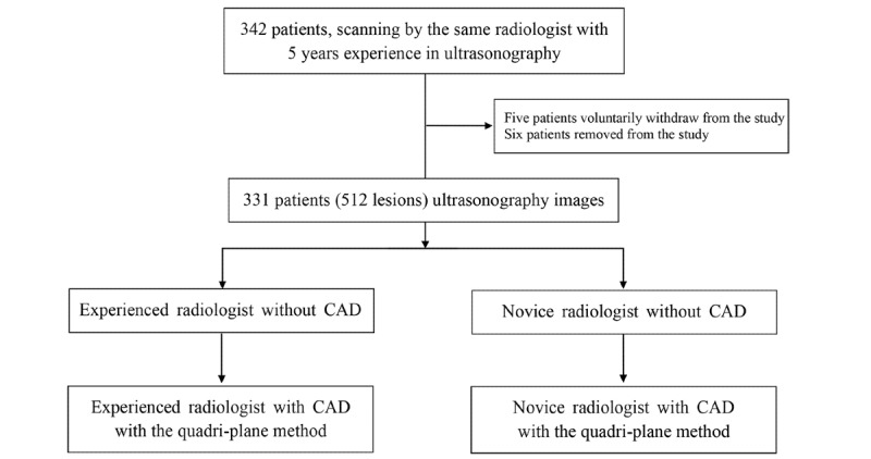
From November 2018 to October 2019, we enrolled patients in the study who had a breast mass as their most prominent symptom. We assigned 2 ultrasound radiologists (with 1 and 5 years of experience, respectively) to read breast ultrasonography images without CAD and then to perform a second reading while applying CAD with the quadri-planes method. We then compared the diagnostic performance of the readers for the 2 readings (without and with CAD). The McNemar test for paired data was used for statistical analysis.

RESULTS

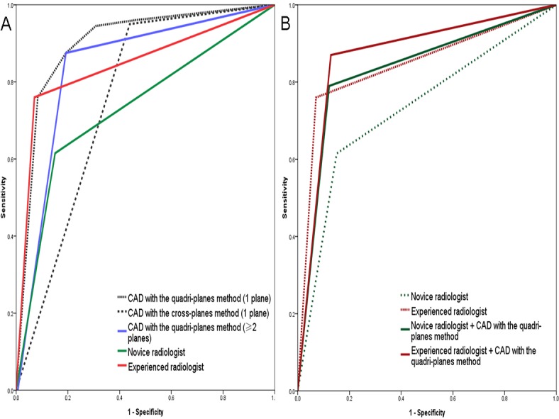
A total of 331 patients were included in this study (mean age 43.88 years, range 17-70, SD 12.10), including 512 lesions (mean diameter 1.85 centimeters, SD 1.19; range 0.26-9.5); 200/512 (39.1%) were malignant, and 312/512 (60.9%) were benign. For CAD, the area under the receiver operating characteristic curve (AUC) improved significantly from 0.76 (95% CI 0.71-0.79) with the cross-planes method to 0.84 (95% CI 0.80-0.88; P<.001) with the quadri-planes method. For the novice reader, the AUC significantly improved from 0.73 (95% CI 0.69-0.78) for the without-CAD mode to 0.83 (95% CI 0.80-0.87; P<.001) for the combined-CAD mode with the quadri-planes method. For the experienced reader, the AUC improved from 0.85 (95% CI 0.81-0.88) to 0.87 (95% CI 0.84-0.91; P=.15). The kappa indicating consistency between the experienced reader and the novice reader for the combined-CAD mode was 0.63. For the novice reader, the sensitivity significantly improved from 60.0% for the without-CAD mode to 79.0% for the combined-CAD mode (P=.004). The specificity, negative predictive value, positive predictive value, and accuracy improved from 84.9% to 87.8% (P=.53), 76.8% to 86.7% (P=.07), 71.9% to 80.6% (P=.13), and 75.2% to 84.4% (P=.12), respectively. For the experienced reader, the sensitivity improved significantly from 76.0% for the without-CAD mode to 87.0% for the combined-CAD mode (P=.045). The NPV and accuracy moderately improved from 85.8% and 86.3% to 91.0% (P=.27) and 87.0% (P=.84), respectively. The specificity and positive predictive value decreased from 87.4% to 81.3% (P=.25) and from 87.2% to 93.0% (P=.16), respectively.

CONCLUSIONS

S-Detect is a feasible diagnostic tool that can improve the sensitivity, accuracy, and AUC of the quadri-planes method for both novice and experienced readers while also improving the specificity for the novice reader. It demonstrates important application value in the clinical diagnosis of breast cancer.

TRIAL REGISTRATION

ChiCTR.org.cn 1800019649; http://www.chictr.org.cn/showproj.aspx?proj=33094.

https://cdn.ncbi.nlm.nih.gov/pmc/blobs/5571/7238092/bd3151dc9309/medinform_v8i5e18251_fig4.jpg
https://cdn.ncbi.nlm.nih.gov/pmc/blobs/5571/7238092/a4d4b8e49bb3/medinform_v8i5e18251_fig1.jpg
https://cdn.ncbi.nlm.nih.gov/pmc/blobs/5571/7238092/c5522f95256f/medinform_v8i5e18251_fig2.jpg
https://cdn.ncbi.nlm.nih.gov/pmc/blobs/5571/7238092/da5fb7529509/medinform_v8i5e18251_fig3.jpg
https://cdn.ncbi.nlm.nih.gov/pmc/blobs/5571/7238092/bd3151dc9309/medinform_v8i5e18251_fig4.jpg
摘要

背景

计算机辅助诊断(CAD)是一种可帮助放射科医生通过超声检查诊断乳腺病变的工具。以往研究表明,CAD有助于降低放射科医生漏诊的发生率。然而,尚未评估使用诊断平面将CAD应用于乳腺病变的最佳方法。

目的

本研究旨在比较不同经验水平的放射科医生在使用CAD结合四平面法检测乳腺肿瘤时的表现。

方法

2018年11月至2019年10月,我们纳入以乳腺肿块为最突出症状的患者进行研究。我们安排2名超声放射科医生(分别具有1年和5年经验)先在不使用CAD的情况下阅读乳腺超声图像,然后在应用CAD结合四平面法的情况下进行二次阅读。然后我们比较了两位读者在两次阅读(不使用CAD和使用CAD)时的诊断表现。配对数据的McNemar检验用于统计分析。

结果

本研究共纳入331例患者(平均年龄43.88岁,范围17 - 70岁,标准差12.10),包括512个病变(平均直径1.85厘米,标准差1.19;范围0.26 - 9.5);200/512(39.1%)为恶性,312/512(60.9%)为良性。对于CAD,受试者操作特征曲线(AUC)下的面积从使用交叉平面法时的0.76(95%可信区间0.71 - 0.79)显著提高到使用四平面法时的0.84(95%可信区间0.80 - 0.88;P <.001)。对于新手读者,AUC从无CAD模式下的0.73(95%可信区间0.69 - 0.78)显著提高到四平面法联合CAD模式下的0.83(95%可信区间0.80 - 0.87;P <.001)。对于经验丰富的读者,AUC从0.85(95%可信区间0.81 - 0.88)提高到0.87(95%可信区间0.84 - 0.91;P = 0.15)。经验丰富的读者和新手读者在联合CAD模式下的一致性kappa值为0.63。对于新手读者,敏感性从无CAD模式下的60.0%显著提高到联合CAD模式下的79.0%(P = 0.004)。特异性、阴性预测值、阳性预测值和准确性分别从84.9%提高到87.8%(P = 0.53)、从76.8%提高到86.7%(P = 0.07)、从71.9%提高到80.6%(P = 0.13)和从75.2%提高到84.4%(P = 0.12)。对于经验丰富的读者,敏感性从无CAD模式下的76.0%显著提高到联合CAD模式下的87.0%(P = 0.045)。阴性预测值和准确性分别从中度从85.8%和86.3%提高到91.0%(P = 0.27)和87.0%(P = 0.84)。特异性和阳性预测值分别从87.4%降至81.3%(P = 0.25)和从87.2%升至93.0%(P = 0.16)。

结论

S - Detect是一种可行的诊断工具,可提高新手和经验丰富读者四平面法的敏感性、准确性和AUC,同时也提高新手读者的特异性。它在乳腺癌临床诊断中显示出重要应用价值。

试验注册

中国临床试验注册中心ChiCTR.org.cn 1800019649;http://www.chictr.org.cn/showproj.aspx?proj = 33094。

相似文献

1
Evaluation of the Quadri-Planes Method in Computer-Aided Diagnosis of Breast Lesions by Ultrasonography: Prospective Single-Center Study.超声检查中四平面法在乳腺病变计算机辅助诊断中的评估:前瞻性单中心研究
JMIR Med Inform. 2020 May 5;8(5):e18251. doi: 10.2196/18251.
2
Performance of Computer-Aided Diagnosis in Ultrasonography for Detection of Breast Lesions Less and More Than 2 cm: Prospective Comparative Study.超声检查中计算机辅助诊断对2厘米及以下和2厘米以上乳腺病变的检测性能:前瞻性比较研究
JMIR Med Inform. 2020 Mar 2;8(3):e16334. doi: 10.2196/16334.
3
Performance and Reading Time of Automated Breast US with or without Computer-aided Detection.自动乳腺超声(ABUS)联合或不联合计算机辅助检测的性能和阅读时间。
Radiology. 2019 Sep;292(3):540-549. doi: 10.1148/radiol.2019181816. Epub 2019 Jun 18.
4
Evaluation of the effect of computer aided diagnosis system on breast ultrasound for inexperienced radiologists in describing and determining breast lesions.评估计算机辅助诊断系统对经验不足的放射科医生在描述和判定乳腺病变的乳腺超声检查中的作用。
Med Ultrason. 2019 Aug 31;21(3):239-245. doi: 10.11152/mu-1889.
5
Accuracy and interpretation time of computer-aided detection among novice and experienced breast MRI readers.计算机辅助检测在新手和有经验的乳腺 MRI 读者中的准确性和解释时间。
AJR Am J Roentgenol. 2013 Jun;200(6):W683-9. doi: 10.2214/AJR.11.8394.
6
Diagnostic Value of Breast Lesions Between Deep Learning-Based Computer-Aided Diagnosis System and Experienced Radiologists: Comparison the Performance Between Symptomatic and Asymptomatic Patients.基于深度学习的计算机辅助诊断系统与经验丰富的放射科医生对乳腺病变的诊断价值:有症状和无症状患者之间的性能比较
Front Oncol. 2020 Jul 7;10:1070. doi: 10.3389/fonc.2020.01070. eCollection 2020.
7
Improving digital breast tomosynthesis reading time: A pilot multi-reader, multi-case study using concurrent Computer-Aided Detection (CAD).提高数字乳腺断层合成阅读时间:使用计算机辅助检测(CAD)的多读者、多病例先导研究。
Eur J Radiol. 2017 Dec;97:83-89. doi: 10.1016/j.ejrad.2017.10.014. Epub 2017 Oct 24.
8
Deep Learning-Based Computer-Aided Diagnosis for Breast Lesion Classification on Ultrasound: A Prospective Multicenter Study of Radiologists Without Breast Ultrasound Expertise.基于深度学习的超声乳腺病变计算机辅助诊断:无乳腺超声专业知识的放射科医师的前瞻性多中心研究。
AJR Am J Roentgenol. 2023 Oct;221(4):450-459. doi: 10.2214/AJR.23.29328. Epub 2023 May 24.
9
Decrease in interpretation time for both novice and experienced readers using a concurrent computer-aided detection system for digital breast tomosynthesis.使用数字乳腺断层合成术的计算机辅助检测系统,可减少新手和有经验读者的解读时间。
Eur Radiol. 2019 May;29(5):2518-2525. doi: 10.1007/s00330-018-5886-0. Epub 2018 Dec 13.
10
The diagnostic performance of ultrasound computer-aided diagnosis system for distinguishing breast masses: a prospective multicenter study.超声计算机辅助诊断系统鉴别乳腺肿块的诊断性能:一项前瞻性多中心研究。
Eur Radiol. 2022 Jun;32(6):4046-4055. doi: 10.1007/s00330-021-08452-1. Epub 2022 Jan 23.

引用本文的文献

1
Whole-lesion-aware network based on freehand ultrasound video for breast cancer assessment: a prospective multicenter study.基于徒手超声视频的全病灶感知网络用于乳腺癌评估:一项前瞻性多中心研究
Cancer Imaging. 2025 Jun 16;25(1):75. doi: 10.1186/s40644-025-00892-y.
2
Diagnostic performance of ultrasound with computer-aided diagnostic system in detecting breast cancer.超声联合计算机辅助诊断系统检测乳腺癌的诊断性能
Heliyon. 2023 Oct 6;9(10):e20712. doi: 10.1016/j.heliyon.2023.e20712. eCollection 2023 Oct.
3
The diagnostic performance of ultrasound computer-aided diagnosis system for distinguishing breast masses: a prospective multicenter study.

本文引用的文献

1
Performance of Computer-Aided Diagnosis in Ultrasonography for Detection of Breast Lesions Less and More Than 2 cm: Prospective Comparative Study.超声检查中计算机辅助诊断对2厘米及以下和2厘米以上乳腺病变的检测性能:前瞻性比较研究
JMIR Med Inform. 2020 Mar 2;8(3):e16334. doi: 10.2196/16334.
2
An investigation of the classification accuracy of a deep learning framework-based computer-aided diagnosis system in different pathological types of breast lesions.基于深度学习框架的计算机辅助诊断系统在不同病理类型乳腺病变中的分类准确性研究。
J Thorac Dis. 2019 Dec;11(12):5023-5031. doi: 10.21037/jtd.2019.12.10.
3
Computer-Aided Diagnosis of Solid Breast Lesions With Ultrasound: Factors Associated With False-negative and False-positive Results.
超声计算机辅助诊断系统鉴别乳腺肿块的诊断性能:一项前瞻性多中心研究。
Eur Radiol. 2022 Jun;32(6):4046-4055. doi: 10.1007/s00330-021-08452-1. Epub 2022 Jan 23.
4
A Human-Algorithm Integration System for Hip Fracture Detection on Plain Radiography: System Development and Validation Study.一种用于在普通X线片上检测髋部骨折的人机算法集成系统:系统开发与验证研究
JMIR Med Inform. 2020 Nov 27;8(11):e19416. doi: 10.2196/19416.
5
An Image-Based Mobile Health App for Postdrainage Monitoring: Usability Study.基于图像的引流后监测移动健康应用:可用性研究。
J Med Internet Res. 2020 Aug 28;22(8):e17686. doi: 10.2196/17686.
计算机辅助诊断超声下实性乳腺病变:假阴性和假阳性结果的相关因素。
J Ultrasound Med. 2019 Dec;38(12):3193-3202. doi: 10.1002/jum.15020. Epub 2019 May 11.
4
Feasibility of computer-assisted diagnosis for breast ultrasound: the results of the diagnostic performance of S-detect from a single center in China.乳腺超声计算机辅助诊断的可行性:中国单中心S-detect诊断性能的结果
Cancer Manag Res. 2019 Jan 23;11:921-930. doi: 10.2147/CMAR.S190966. eCollection 2019.
5
Breast Cancer Treatment.乳腺癌治疗
JAMA. 2019 Jan 22;321(3):316. doi: 10.1001/jama.2018.20751.
6
Cancer statistics, 2019.癌症统计数据,2019 年。
CA Cancer J Clin. 2019 Jan;69(1):7-34. doi: 10.3322/caac.21551. Epub 2019 Jan 8.
7
Computer-Aided Diagnosis Based on Convolutional Neural Network System for Colorectal Polyp Classification: Preliminary Experience.基于卷积神经网络系统的计算机辅助诊断用于结直肠息肉分类:初步经验
Oncology. 2017;93 Suppl 1:30-34. doi: 10.1159/000481227. Epub 2017 Dec 20.
8
Ultrasound Imaging Technologies for Breast Cancer Detection and Management: A Review.用于乳腺癌检测与管理的超声成像技术:综述
Ultrasound Med Biol. 2018 Jan;44(1):37-70. doi: 10.1016/j.ultrasmedbio.2017.09.012. Epub 2017 Oct 26.
9
Application of computer-aided diagnosis in breast ultrasound interpretation: improvements in diagnostic performance according to reader experience.计算机辅助诊断在乳腺超声解读中的应用:根据阅片者经验的诊断性能改善
Ultrasonography. 2018 Jul;37(3):217-225. doi: 10.14366/usg.17046. Epub 2017 Aug 14.
10
Application of Computer-Aided Diagnosis on Breast Ultrasonography: Evaluation of Diagnostic Performances and Agreement of Radiologists According to Different Levels of Experience.计算机辅助诊断在乳腺超声检查中的应用:根据不同经验水平评估诊断性能及放射科医生之间的一致性
J Ultrasound Med. 2018 Jan;37(1):209-216. doi: 10.1002/jum.14332. Epub 2017 Aug 1.